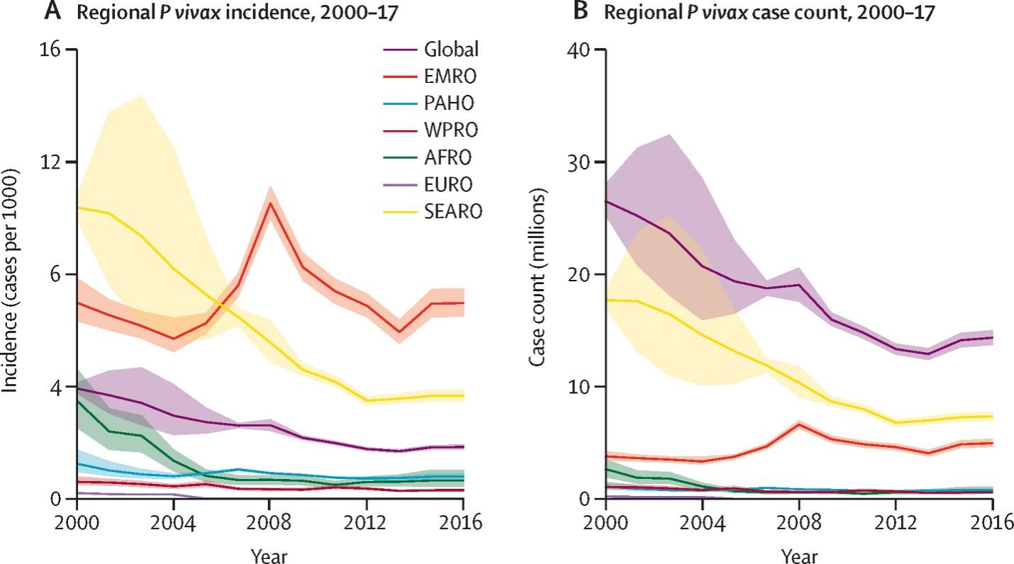
In these logs, we take a deep analysis of the various microorganisms and outline them to regular people who are interested in understanding various aspects related to treatment and symptoms for the microorganisms. Today, we will take a look into the Plasmodium Vivax eukaryote which has in the recent past been making headlines as it causes the common malaria disease (Bourgard et al. 34). This blog aims at analyzing the microorganism on aspects such as its scientific name, eternal structure and functions and the internal structures and functions. Currently, there has been a growing number of cases related to the malaria disease, which is assumed to be the leading cause of death around the globe. It has been terrifying reading various headline blogs, and this is what has prompted the writing of this blog article.

So What the Heck is Plasmodium Vivax?
Classification?
The Malaria Parasite scientifically, known as Plasmodium Vivax, is a human pathogen and a protozoal parasite that has widely and frequently been thought as the recurring cause of malaria. Plasmodium Vivax is also thought to be the deadliest of all the five human malaria parasites despite being lesser virulent than Plasmodium falciparum (Adams et al. 25585). This is based on its cause of severe diseases and even death that is caused by splenomegaly (a pathologically enlarged spleen).
Plasmodium Vivax is believed to have originated from Asia and is currently found mainly in Latin America and various parts of Africa. At least 71 species of mosquitoes are known to carry P. vivax. As far north as Finland, vivax vectors are thriving in moderate conditions (Markus 1765). Indoor insecticides and bed nets may not be as efficient because some prefer to bite during the daytime. Insecticide resistance has yet to be assessed in some important vector species that have not yet been cultivated in the lab for further investigation.
External Structures and Functions?
Compared to P. falciparum, Plasmodium Vivax can remain in your liver for up to three years because of its milder symptoms. P. vivax is not the most lethal kind of malaria. In addition to being a human disease, it is a protozoan parasite. Let us highlight some interesting facts about Plasmodium vivax:
- A trophozoite is the term used to describe the parasite’s mature form.
- An amoeboid, the trophozoite is uninucleated.
- The female Anopheles mosquitoes are the carriers of P. vivax.
- This parasite is not present in the Anopheles males.
The female Anopheles mosquitoes are the ones that carry and transfer the parasite to the human host. They are the actual conduit to the host. Both mosquitoes have a separate life cycle that can’t be compared to the other (Adams et al. 25585). The female acts as a vector for the parasite, which she then transfers to the human host.
Rings: Plasmodium vivax has a signet ring shape and a big cytoplasm that contains a huge chromatin, making it unique. They take on an amoebic appearance as they get older. The parasite’s ring form has a diameter around one-third that of human red blood cells.
Trophozoites: Plasmodium vivax trophozoites have a yellowish-brown pigment (hematin) in their cytoplasm, amoeboid cytoplasm and big chromatin spots. It functions as a vegetative form for reproduction.
Gametocytes: Plasmodium vivax gametocytes have a more defined form than trophozoites because they are spherical and oval in shape (Bourgard et al. 34). Infected red blood cells are characterized by a high concentration of brown pigments that spread throughout the cells.
Schizonts: There are 12 to 24 merozoites in the schizonts of Plasmodium vivax, which are big enough to fill the cell (red cell). Additionally, after staining, a yellowish-brown pigment can be detected on the cells.
Internal Structures and Functions?
Bulbous rhoptries: They include proteins that aid in the invasion and modification of the host cell.
Micronemes: Micronemes, which are parasite proteins that aid in movement and host cell attachment, are located next to the rhoptries.
Dense Granules: Dense granules, secretory vesicles seen throughout the parasite, contain proteins that aid in the modification of the parasitophorous vacuole.
Single Large Mitochondrion: Aids in the coordination of its division with the larger plasmodium viva cell
Apicoplast: It is a large membrane bound organelle of endosymbiotic origin which play a role in parasitic metabolism
Ribosomes: Plasmodium Vivax have a structurally distinct, stage specific ribosomes. A further purpose for these nucleotides was to track the turnover of insect ribosomal proteins during parasite growth.

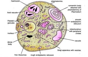
In the picture above, there are various internal structures found in a typical plasmodium vivax cell. Structures illustrated include: the nucleus, mitochondrion, vacuole, Golgi apparatus, rough endoplasmic reticulum, ribosomes, smooth endoplasmic reticulum, food vacuoles, plasmallema and haemozoin.
Works Cited
Adams et al. “The Biology of Plasmodium Vivax.” Cold Spring Harbor perspectives in medicine, 2017.
Ollomo, Gilbert. “Plasmodium vivax-like genome sequences shed new insights into Plasmodium vivax biology and evolution.” PLOS Biology, Web.
Bourgard, Catarina, et al. “Plasmodium Vivax Biology: Insights Provided by Genomics, Transcriptomics and Proteomics.” Frontiers in cellular and infection microbiology 8 (2018): 34. Web.
Markus, Miles B. “Biological Concepts in Recurrent Plasmodium Vivax Malaria.” Parasitology, 2018. Web.