Introduction
Compliance with safety precautions is imperative for health workers in the radiology department as they may be at particular risk if they are not appropriately equipped. This necessitates the creation of a particular environment in which there will be conditions for staff and patients to be safe. Installation of radiation filtering layers is a mandatory important factor that can affect the quality of health of both doctors and clients. Satisfying the need for standard precautions and quality medical care is imperative and can be achieved by installing new equipment.
Scope of Work
A needs assessment is a significant step that must be carried out to understand all the currently existing gaps that must be addressed. In this context, there is a need to provide sufficient protection for clinic staff and clients. This is justified by the need to protect the health and well-being of staff to ensure the best working conditions.
Several solutions were selected as the necessary equipment; one is radiation-absorbing filters that can provide at least a 2.5 mm Half Value Layer (Sherer et al., 2014). In addition to this element, shielding the room fully with lead to reflect radiation will be necessary. This may have a more comprehensive effect by providing radiation absorption. In addition, an essential element is the placement of indicators that should notify that the radiation is on.
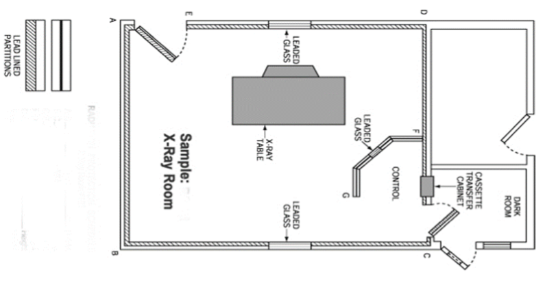
The design and layout of the premises are imperative to ensure preventive protection from radiation exposure. An example of providing a reliable, efficient, and safe environment is shown in Figure 1. The room is designed to separate all empty spaces by lead floors (Lux Construction, Inc., 2023). In addition, the space is filled in such a way that it is close to the external clinic on all sides. With the help of such an overlap, achieving the maximum-security effect in combination with special filters will be possible. Installation and testing of the central X-ray equipment and protective layers should occur in stages by the established plan (Sherer et al., 2014).
Thus, the first point is to check the correct placement. After this, the placement of lead plates on the walls should begin. The X-ray machine will also need to install the filters discussed above. The last point is the equipment calibration for testing and measuring radiation levels. This process can be supported by obtaining quality information from evidence-based sources (University of Doha, n. d.). In addition, a necessary aspect is the involvement of specialists who could improve the process and make it as high quality as possible.
Timeline
Space planning will begin on November 25, 2023. Thus, the first stage of the final project will be completed in three days. After this, purchasing equipment through suppliers is imperative, which, together with delivery, will take five days and will be completed on December 3, 2023. Next, the installation and testing process will last until December 10, 2023. The total time spent on creating the converted X-ray room was 15 days.
Budget
The cost of purchasing equipment is the most significant element in the installation system of an x-ray room. To fully provide everything, it will be necessary to allocate 50,000 US dollars for an X-ray station to take pictures. After this, one will need to add the purchase of lead sheets, which can cost $4,000. General equipment installation and finishing services will cost $7,000. Thus, the total cost of equipping the x-ray room would be $61,000.
Conclusion
The arrangement of the X-ray room must be carried out according to all norms and standards in the field of ensuring the health of medical staff and clinic clients. Creating a quality design to determine schedules and possible necessary design interventions is a necessary step in a long production process. Providing protective measures should be one of the main aspects that need to be taken care of during the planning and design of an X-ray room.
References
Lux Construction, Inc. (2023). X-ray/radiology room design for hospital [checklist]. Web.
Sherer, M. A., Visconti, P., Ritenour, R. & Heynes, K. (2014). Radiation protection in medical radiography (7th ed.). Mosby.
University of Doha. (n. d.). CLO5: Equipment Specifications and Shielding Guidelines [PowerPoint slides]. AHMR 1203.