Nature has made lots of molecule and enzyme substrates which are important in various reaction paths.
Yeast alcohol dehydrogenase refers to a group of enzymes that are found in yeast and have a widespread application in the beer and wine industry where they facilitate the process of fermentation. Their basic functioning reaction involves the facilitation of conversion of alcohols to aldehydes or ketones and vice versa. This process is usually accompanied by a reduction of nicotinamide adenine dinucleotide (NAD+ to NADH). They are very important in the human and animal body functioning as they facilitate the breakdown of toxic alcohols into less toxic compounds and they also enhance the production of important metabolites that are used in various biosynthetic processes (Branden 1975, p.134).
Characteristically, alcohol dehydrogenases are a composition of several isozymes which catalyze the oxidation of alcohols to aldehydes and ketones. Primary alcohols are oxidized into aldehydes while secondary alcohols are oxidized to ketones. The process is more expressed in mammals as a redox reaction involving the reduction of the coenzyme nicotinamide adenine dinucleotide (NAD+) and the oxidation of the alcohol. Alcohol dehydrogenase is expressed as a dimer with a mass of 80kDa (Eklund et al., 1976).
The protein structure shows evidence of uneven distribution of the residual proteins. This is characteristically portrayed in arginine in the T-terminal half and tryptophan in the N-terminal half. The same is observed at a higher extent in other residues including cysteine, histidine, and proline. These residues are present in large amounts or rather are found as an over-expression in the N-terminal half (Rossmann 1975, p.78).
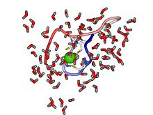
Yeast alcohol dehydrogenase has a structural zinc site where the zinc ion plays a vital role in fortifying protein stability. These structural sites are revealed in crystallographic structures by classical molecular techniques and quantum chemistry. The structure portrays four Cysteine ligands which are found in close proximity to each other. They include Cys97, Cys100, Cys103 and Cys111. These ligands are located around the zinc ion in asymmetric tetrahedron orientation. This arrangement is enhanced by electrostatic forces and additional covalence binding forces.
It has also been shown that the protein has non-random sequence patterns. In yeast alcohol dehydrogenase, approximately 60 percent of all valine residues are located adjacent to other branched-chain residues and this usually occurs at five different positions, Val-Val or Val-Val-Val. Other repetitive residuals are also seen to occur with many triple-residual sequences being similar. In this regard, the following identical sequences are common : 21-23 and 225-227; 35-37 and 243-245; 36-38 and 78-80; 45-47 and 143-145; 72-74, (319)-(321) and (337)-(339); 88-90 and 154-156;127-129and188-190;133-135and186-188;202-204 and 214-216; 227-229 and (290)-(292).
It can also be identified that in some regions of the protein, the structure is defined by some small residues or proline that is, at positions 24-28 and 55-61. Also, hydrophobic regions are evident in the structure of yeast dehydrogenase. Long segments which occur without any charged residues occur and can be seen at positions 172-190, 261-274 and at positions 287-297. This orientation of small residues and proline is particularly important during the unfolding process. The same has also been attributed to the tertiary structure that the protein may at times be seen to assume. The presence of glycine and some repetitive structures are particularly important in determining the evolutionary relationships between different proteins. The presence of hydrophobic areas in the protein is the reason why some portions of this protein are insoluble in water. When the protein is digested by staphylococcal protease, the resulting large precipitates of trypsin that correspond to regions 261-298 of the protein are completely insoluble in water. It is for this reason that the T8 forms a precipitate in tubes after its consequent separation through exclusion chromatography. (Branden 1975, p.134).
These hydrophobic areas are important as they may explain other properties that may be associated with the protein. They are especially important in defining the nature of the physicochemical properties of the yeast dehydrogenase enzyme. Gel electrophoresis of yeast alcohol dehydrogenase and horse alcohol dehydrogenase suggests that the carboxy-methylated subunits of the horse protein are slightly small than those of the yeast protein. This is completely contrary to what is provided by native protein sequences that suggest the opposite. The results of gel electrophoresis have been strengthened by the fact that isolated subunits through maleylation show similar evidence. This particular characteristic has been attributed to reversals that occur due to differences in the distribution of hydrophobic residues in these two enzymes. These differences have also been said to be related to the quaternary structure and thus to subunit interactions (Veillon, 1975)
From the mentioned differences, it has been concluded that the yeast alcohol dehydrogenase can bind more sodium dedocylsulphate. It has also been said that different quaternary structures affect the method of determining the relative molecular weights of the monomers (Rossmann 1975, p.78).
References
Branden, C.-I., Jornvall, H., Eklund, H. & Furugren, B., 1975. Alcohol Dehydrogense, New York: Academic Press.
Eklund, H., Branden, C.-I. & Jornvall, H., 1976. Alcohol Dehydrogenase, Journal of Molecular Biology.
Jornvall, H. ,1973. Differences between Alcohol Dehydrogenases. PNAS
Jornvall, H., 1974. Alcohol and Aldehyde Metabolizing Systems, New York: Academic Press.
Leskovac, V.; Trivic, S.; Peričin, D., 2002. The three zinc-containing alcohol dehydrogenases from baker’s yeast, Saccharomyces cerevisiae, FEMS Yeast Research 2.
Rossmann et al., 1975. Structure of Alcohol Dehydrogenase,New York: Academic Press.
Sund, H. & Theorell, H., 1963. Alcohol Dehydrogenase, New York: Academic press.
Veillon, C. & Sytkowski, A. J., 1975. The Intrinsic Zinc Atoms of Yeast Alcohol Dehydrogenase, Biochemical and Biophysical Research Communications.