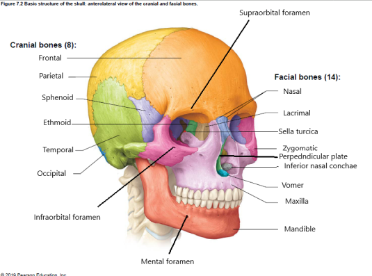
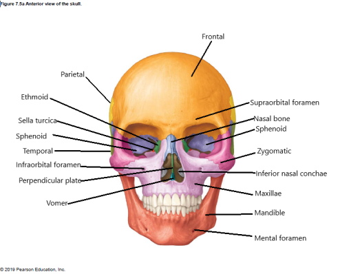
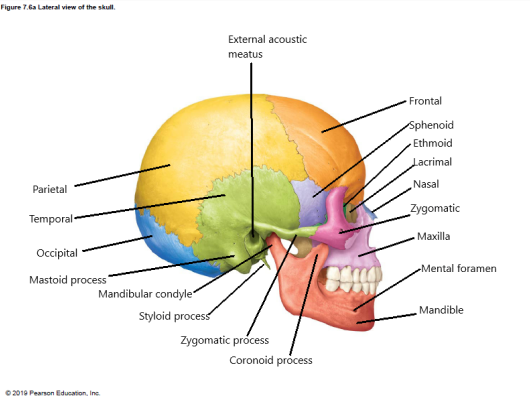
Skull – consists of cranial and facial bones
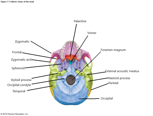
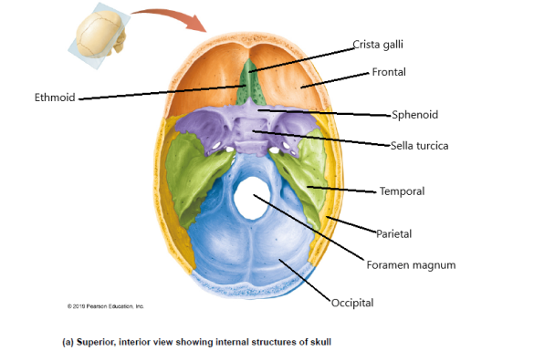
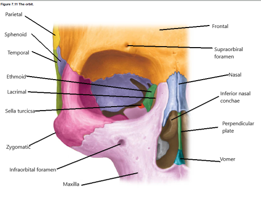
Cranial bones – these contribute to the cranium, which encloses & protects the brain; note they are separated by jagged boundaries called sutures; there are 8 cranial bones (note there is a right & left parietal and a right & left temporal; (label the below bones in figures 7.2, 7.5A, 7.6a, 7.7, 7.9a, 7.11)
Parts of the mandible:
Condylar process – Each one of these *articulates with a temporal bone.
Vertebral Column
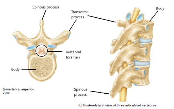
General structure of a vertebra (you will find the following on all vertebrae. (label the below structures in figure 7.18)
- Body
- Vertebral foramen
- Transverse process
- Spinous process
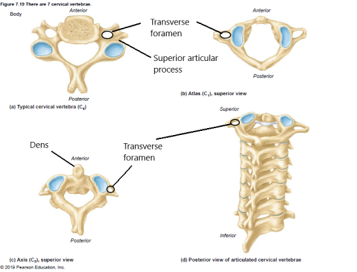
Specializations of cervical vertebrae include (label the below specializations in figure 7.19)
- Transverse foramen; all cervical vertebrae (and only cervical vertebrae) have this pair of openings in their transverse processes
- The atlas is the first vertebra, which possesses unique features because of its *articulation with the occiput of the head:
- Superior articular processes – These are shaped differently from those of any other vertebra to articulate with the contours of the occipital condyles of the skull.
- The axis is the second vertebra, which has a unique structure to permit it to rotate the atlas and skull:
- The dens *articulates with the atlas at the inner surface of the anterior arch
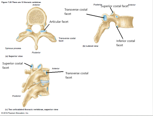
Specializations of thoracic vertebrae (label the below specializations in figure 7.20)
Costal facets – All thoracic vertebrae (and only thoracic vertebrae) have costal facets (“costal” means “rib”). These special articular processes are *for articulation with the head of the rib.
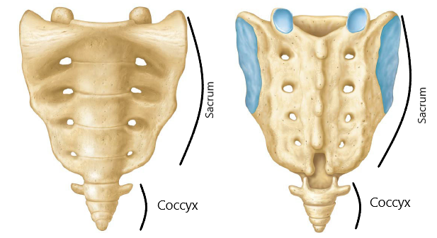
Sacrum and coccyx – During development, the sacrum is formed from the fusion of five separate fetal vertebrae; the coccyx is the tiny, inferior bone commonly known as the “tail” bone. (label the sacrum and coccyx in figure 7.22)
Thorax
Consists of thoracic vertebrae, sternum, and ribs; label the below bones, parts, AND MARKINGS on figures 7.24 and 7.25.
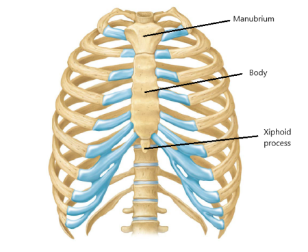
Sternum – the “breast bone” consists of 3 parts:
- Manubrium – superior, heart-shaped
- Body – longest part
- Xiphoid process – smallest part; occasionally absent, which indicates a younger person in whom ossification is not complete
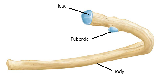
Ribs – all ribs have the following structure:
- Head – bulbous vertebral end, which *articulates with the body of the thoracic vertebrae.
- Tubercle – knuckle-like projection just beyond the head, which *articulates with the transverse process of the more posterior thoracic vertebrae.
- Body – the remainder of the rib’s length.
Appendicular Skeleton
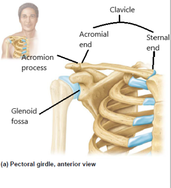
The Pectoral Girdle: Label the Below Bones, Parts, and Markings on Figures 7.27 and 7.28
- Clavicle – There are two of these “collar bones”.
- Sternal end – more blunt end, shaped like a pyramid, for *articulation with the sternum.
- Acromial end – The opposite end is more flattened, and *articulates with the acromion process of the scapula (you’ll identify this bone next).
- Scapula – This is the “shoulder bone”.
- Spine – fin-like projections run across the entire posterior side of the bone.
- Acromion process – roughened projection of the spine; *articulates with the acromial end of the scapula.
- Glenoid fossa – shallow depression for *articulation with the head of the humerus.
The Upper Limb
Label the Below Bones, Parts, and Markings on Figures 7.29 and 7.30 & 7.32.
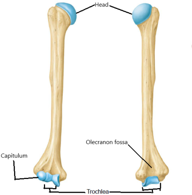
- Humerus – This is the upper arm bone.
- Head – This is the smooth spherical projection which *articulates with the scapula at the glenoid fossa.
- Olecranon fossa – This depression is in the posterior side of the distal end of the bone. It is for the reception of the “elbow”; it *articulates with the olecranon process of the ulna (studied shortly).
- Trochlea – This spool-shaped process is for *articulation with the ulna (in conjunction with the olecranon fossa).
- Capitulum – This rounded process is for *articulation with the radius (studied next); it is just to the side of the trochlea.
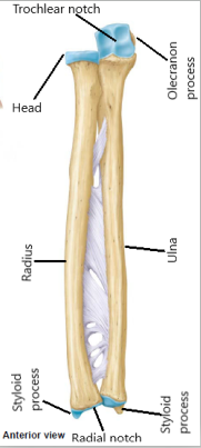
- Radius – In anatomical position, this is the lateral forearm bone.
- Head – This is the proximal, flat, round process. It *articulates with the capitulum of the humerus and laterally with the ulna.
- Styloid process – This lateral projection from the distal end is the bump that can be felt on your arm just before the wrist.
- Ulna – This is the medial forearm bone.
- Olecranon – This is the knob-like projection of the proximal end; the “elbow”.
- Radial notch – This lateral smooth structure *articulates with the head of the radius.
- Trochlear notch – This “crescent moon” structure *articulates with the trochlea of the humerus.
- Styloid process – Like its counterpart on the radius, but medial. This bump can be felt on the posterior forearm just proximal to the wrist.
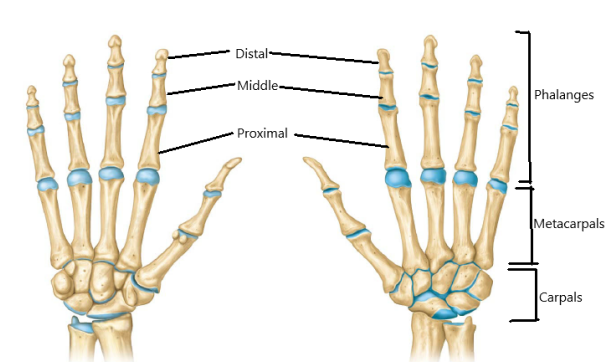
- Carpals – There are 8 of these wrist bones, whose individual names you will NOT be required to learn.
- Metacarpals – “Meta” means middle. There are 5 of these palm bones.
- Phalanges (singular is phalanx) – There are a total of 14 of these “finger bones”.
- Proximal – Each of the five digits will have this as the one which articulates with its respective metacarpal
- Middle – there are only four of these, for digits 2-5.
- Distal – each digit will have this as the final segment.
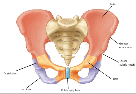
The Pelvic Girdle
Label the Below Bones, Parts, and Markings on Figures 7.34 and 7.35.
Hip (coxal) bone – Two of these complicated bones comprise the pelvic girdle.
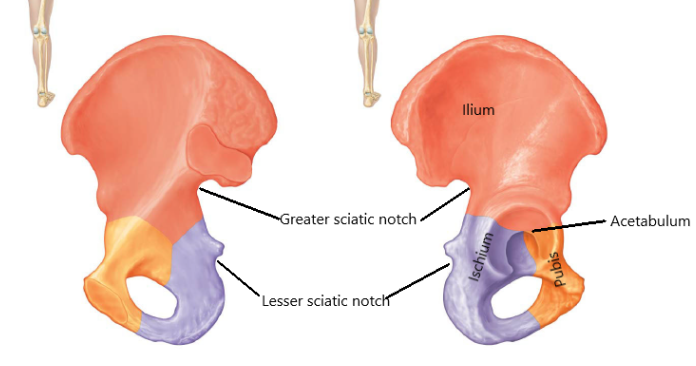
- Ilium – the most superior region articulates with the sacrum
- Sciatic notch – This is the very large notch in the posterior, inferior pelvic border. The sciatic nerve (the longest nerve in the body) passes through this notch.
- Ischium – generally the inferior and posterior pelvic region
- Pubis – inferior and anterior pelvic region
- Pubic symphysis –the surface where the right and left pubic bones join
- The following is not associated with any one of the three pelvic regions
- Acetabulum – this large crater on the lateral side *articulates with the head of the humerus (which you will study next).
The Lower Limb
Label the Below Bones, Parts, and Markings on Figures 7.37 and 7.38 & 7.39.
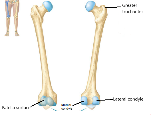
Femur – the thigh bones:
- Head – articulates with the acetabulum of the pelvic bone.
- Greater trochanter – this large roughened projection is opposite the head
- Lateral condyle – rounded knob-like projection from the distal end; *articulates with the lateral condyle of the tibia (which you will study shortly)
- Medial condyle – same as the lateral but positioned medially; *articulates with the medial condyle of the tibia (which you will study shortly) [note: you must orient your bone (right or left) before you can determine which condyle is lateral & which is medial]
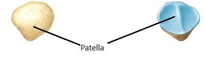
- Patellar surface – this is a smooth, shallow fossa on the anterior side for *articulation with patella or kneecap.
Patella – “kneecap”; The patella *articulates with the patellar surface of the femur.
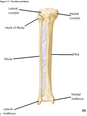
Tibia – “shin bone”
- Lateral condyle – outer smooth rounded half of the proximal end for *articulation with the lateral condyle of the femur. The lateral condyle also *articulates with the head of the fibula (studied next).
- Medial condyle – inner half of the proximal end for *articulation with the medial condyle of the femur.
- Medial malleolus – this distal projection forms the inner ankle. It *articulates with the talus of the ankle (studied shortly).
Fibula – the thinner, more lateral bone of the lower leg
- Head – this is the more rounded proximal end that does not articulate with the femur. Rather, the head of the fibula *articulates with the lateral condyle of the tibia.
- Lateral malleolus – this distal projection forms the outer ankle. Like the tibia, it also *articulates with the talus of the ankle (studied next).
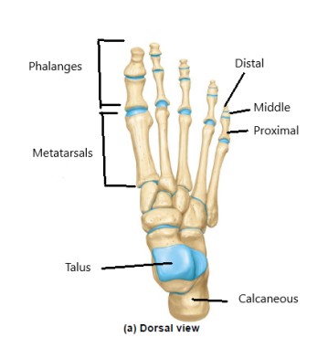
- Tarsals – These 7 bones are equivalent to the carpals. You do not have to know the names of all 7 but you should know:
- Talus – most superior tarsal bone; *articulates with both the calcaneus and the navicular
- Calcaneus – “heel bone”; the largest and strongest tarsal
- Metatarsals – These are equivalent to the metacarpals (“meta” means middle).
- Phalanges (singular is phalanx) – there are fourteen, just as in the hand; remember to give location & numbest
- Proximal
- Middle – not present in great toe
- Distal