Abstract
Leprosy is a highly contagious disease that is caused by M. leprae. The disease affects the cooler parts of the body, especially acral parts of the body. Leprosy is endemic and mainly found in low socio-economic communities across the globe. Leprosy evolves from a local to a general form of the disease as it changes from TT to LL. The host’s T-cell immunity determines how the disease manifests, with strong T-cell immunity restricting the growth of the bacteria greatly. However, the nerve damage, in this case, is usually severe, resulting in a TT type of disease. The long incubation period in leprosy may complicate diagnosis. Leprosy can be cured completely using multidrug bactericidal treatment. Registered nurses should help in advising and educating patients on symptoms, treatment, and prevention of leprosy. These efforts should also be focused on persons living or have lived in endemic areas and those living or have lived with infected persons.
Epidemiology
Leprosy is a systemic disease that is chronic in nature and highly contagious. Leprosy is also known as Hansen’s disease. It is a granulomatous illness whose etiologic agent is Mycobacterium leprae, an acid-fast bacillus. The disease involves the cooler parts of the body, including the mucosal part of the upper airways, the skin, and the neural network (Yuchua-Guillen & Dofitas, 2012).
Clinical leprosy affects not more than 5% of the people who get exposed to M. leprae. Subclinical infections in endemic areas range from 1.7-35% of persons exposed to the causative agent (Worobec, 2009). There is no specific mode of leprosy transmission, but a number of transmission modes have been enumerated. It is possible to acquire leprosy infection after exposure to the bacilli in mucus or oral secretions. Skin contact with an infected person can transmit the bacteria. The disease can also be transmitted congenitally. Contaminated needles, especially needles used in dermal procedures like tattooing, can also pass the infection. It is also suggested that exposure to vectors like insects or infected soil can transmit the disease.
Brazil has the highest cases of Hansen’s disease, with up to 80% of all the cases reported in the Americas coming from Brazil (Worobec, 2009). The disease is largely associated with low socioeconomic status such that most cases of leprosy are reported among persons with low education levels, food shortage, bathing in open water, and living in mud floored houses. These factors, which portray poverty, increase the chances of transmitting the disease.
The World Health Organization (WHO) launched a program in 1991 to eliminate leprosy globally. Despite having succeeded in wiping away leprosy in 118 out of 122 target countries, the disease is still present in all the continents. India leads in cases of leprosy ever registered. Pockets of leprosy cases are still registered in countries such as D.R. Congo, Greece, Mozambique, Nepal, Brazil, Spain, the US states like Louisiana and Texas, and Angola among other countries across the continents. Some countries such as Norway have already registered almost 100% elimination of leprosy (Worobec, 2009).
Pathophysiology
Leprosy patients have lepra reactions that lead to serious morbidity, especially if the diagnosis is not made early. Class IV hypersensitivity is the first reaction that is characterized by a high immune response from T-cells. The patient downgrades to the point where lepromatous Hansen’s disease shows. Foot drop and facial palsy are evident as painful neuritis. Oedema is also a hallmark sign. Class II hypersensitivity is also called the erythema nodosum leprosum. The patient develops erythmatous nodules that are tender and may ulcerate. The patient is febrile at this stage, with glomeluranephritis and orchitis, among other forms of inflammatory reactions, appearing (Sheetal & Arvind, 2009).
Classification and Clinical Presentation
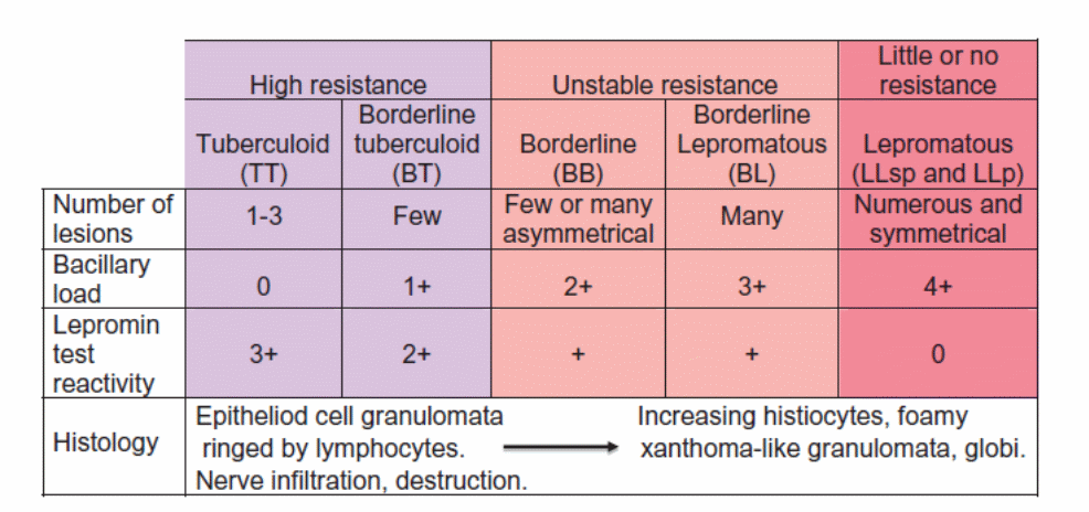
Leprosy has a long incubation period, ranging from 3 months to 40 years. Nevertheless, the typical incubation period is between 5 and 7 years. A person’s T-cell immunity response is a determinant factor in the type of leprosy one acquires and the level of progression. In this regard, there are four clinical spectra for leprosy: The ‘pauci-bacillary’ immunocompetent polar tuberculoid (TT) spectrum, the anergic ‘multibacillary’ polar lepromatous (LL) spectrum, the intermediate spectrum (I), and the borderline spectra (BT and BL) (Worobec, 2009).
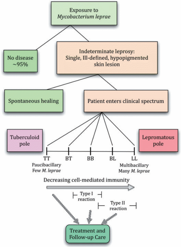
A strong T-cell immune response results in the TT type of leprosy. A low T-cell immunity is characterized by bacterial dissemination in a wide area. This results in the LL stage of the disease. Intermediate T-cell immunity results in the borderline subsets of the disease (Sheetal & Arvind, 2009).
Polar Tuberculoid (TT)
This form of leprosy is common amongst people who have a highly resistant T-cell immunity, but the resistance is not complete. Patients suffering from TT form of leprosy report neural numbness, muscle weakening, and paralysis. The fourth and fifth finger may present with weakness. Dry skin lesions are present singly, or in 2-3 flat patches. The lesions are present in cooler parts of the body. Most of the lesions appear as hypopigmented and hardly do they appear erythmatous (Yuchua-Guillen & Dofitas, 2012). The lesions are largely insensitive on the surface, except in areas where nerve supply in the dermis is rich. Facial lesions lead to inability to sense temperature changes. Histologically, the skin shows minute tuberculoid granulomas.
Polar lepromatous (LL)
The polar lepromatous (LL) stage of the diseases begins from the intermediate form of leprosy for people who have low resistance. It can also start as a deteriorating progression in borderline lepromatous (BL). The disease infiltrates the dermis in a nodular or a non-nodular manner. There is symmetrical distribution of minute hypopigmented macules (Yuchua-Guillen & Dofitas, 2012). Lepromatous infiltrates can also be seen as diffuse or in nodular form. Sensory loss on the macules and sweating loss are not seen, but sweating is later lost. Hair loss is seen on the eyebrows and may later progress to the eyelashes and other body parts. However, there is no hair loss on the head. Nodular infiltration is also seen on knees, earlobes, and hands among other acral parts. Enlargement of the nodules causes destroys underlying structures, such as the nasal cartilage. This eventually causes nose deformity. Loss of teeth results when the alveolar ridge is fully eroded (Worobec, 2009).
Borderline Tuberculoid (BT)
The clinical findings present in BT phase are almost the same as those of TT. However, skin lesions are more and every large lesion has satellite lesions surrounding it. Lesions that occur solely are larger than those that occur together.
Borderline Lepromatous (BL)
It is common to find a first time lesion on the skin in BT. Innumerable symmetrical lesions that are composed of macules, nodules, and plaques are present. Up to 33% of the cases have annular lesions that have a diffuse outer border. The inner border is clearly demarcated. Sensory loss is not experienced in cases where there a multiple lesions. The smaller lesions are more outstanding in such cases. There is symmetrical nerve involvement later. Some of the most pronounced findings in PL, such as leonine facies and saddle nose, are not present in BL (Worobec, 2009).
Borderline (BB)
There are more lesions on the skin in the BB phase. There is asymmetrical distribution of the lesions, although the lesions are not well defined. Instead, smaller lesions are common in satellite form.
There are some variant clinical presentations observed in leprosy cases. For instance, there is a histoid form of multibacillary Hansen’s disease that presents as nodules and papules located on the body’s midsection. There is also the pure neuritic leprosy variant that shows as numerous peripheral nerve involvements in an asymmetrical pattern. Sensory loss on the trunk nerves is common, with motor deficit occurring in some cases. Nevertheless, there are no skin lesions in this form of disease. Pure neuritic leprosy is common in some Africa, Nepal, and India. Involvement of the 7th nerve affects the eyes to an extent that further involvement of ocular structures leads to blindness (Worobec, 2009).
Advanced form of the diseases causes bateremia that affects vital body organs such as liver and lymph nodes. ‘Beautiful leprosy’, or the diffuse lepromatous leprosy, is also another variant of leprosy that causes smoothening of the skin in its initial stages. This variant does not cause nodules; instead, it makes patients look younger. However, madarosis and telangiectasia are noted. It is more common along the Caribbean (Yuchua-Guillen & Dofitas, 2012).
Diagnosis
The long incubation period in leprosy complicates accuracy in diagnosis. Diagnosis begins with being aware that persons who have sensory loss and skin lesions and have stayed in areas that are considered to be endemic may be having Hansen’s disease. Persons who have above symptoms and have lived with colleagues who have the disease are also likely to have leprosy. Although physical appearance of lesions and patient’s description of memory loss are used in establishing diagnosis, histological findings are the best standard for making accurate diagnosis (Sheetal & Arvind, 2009).
Presence of M. leprae and granulomatous disease is determined using staining methods; that is, Fine and H&E stains. The extent of disease is determined using smears of slit skin to test for acid fast bacteria. A lesion that heals by itself is usually the first clinical symptom of intermediate leprosy. The various stages of Hansen’s disease, TT, BT, BB, BL, and PL, show different characteristics as described above. Intermediate leprosy is diagnosed by identifying hypopigmented patches (Worobec, 2009). The ability to sense light touch and differentiate between hot and warm temperatures acts as differential diagnosis in identifying persons who have been in endemic regions and those who have lived with family members who have, or had leprosy.
Treatment
It is possible to cure leprosy completely, especially if diagnosis is made at an early stage. Leprosy was traditionally treated with dapsone, a weak baceteriocidal agent. However, multidrug treatment consisting of rifampin, clofazimine and dapsone was introduced by the World Health Organization (WHO) in 1982. The most effective drug among the three in killing the bacteria is rifampin. Patients who are undergoing rifampin treatment become non-infectious a few days after starting treatment (Worobec, 2009).
The essence of using a combination of 3 drugs is to reduce chances of resistance to rifampin. Dapsone and clofamizine act synergistically since both are weakly bactericidal. Other antibacterial agents such as ofloxacin and clarithromycin are also effective in treating leprosy. Multibacillary disease is advisably treated in 1 year. Paucibacillary patients who present with one skin lesion are advised to take a single-dose composed of rifampin, ofloxacin and minocyclin. However, the WHO recommends that treatment should be extended to increase chances of complete cure (Sheetal & Arvind, 2009).
It is advisable to conduct follow ups on patients after treatment, possibly on an annual basis, to prevent possible relapse. Educating patients about the disease goes a long way in complementing antimycobacterial treatment. Other advisable modes of treatment including offering social support, treating reactions, taking care of nerve damage, and providing occupational and physical therapy. Family members of the patient should also be empowered with information about clinical signs that foretell leprosy (Worobec, 2009).
The Roles of Registered Nurses
Registered nurses (RN) have a critical role to play in diagnosing, treating, and preventing infection and spread of Hansen’s disease. Registered nurses should be in the forefront in educating communities that live in endemic areas about symptoms of leprosy and helping them seek medical attention to establish diagnosis. It is also the role of RNs to educate leprosy patients, their families, and persons living in endemic areas on how to avoid M. leprae infections. RN should also help in setting up social support centers for persons suffering from leprosy.
References
Sheetal, S., & Arvind, C. (2009). Lest we forget Hansen’s disease (leprosy): an unusual presentation with an acute onset of inflammatory polyarthritis and the rheumatology experience. International Journal of Rheumatic Diseases, 12, 64-69.
Worobec, S. P. (2009). Treatment of leprosy/Hansen’s disease in the early 21st century. Dermatologic Therapy, 22, 518-537.
Yuchua-Guillen, A., & Dofitas, B. L. (2012). Atypical Hansen’s disease presenting as florid verrucous plaques on the lower extremities: a case report. International Journal of Dermatology, 51, 697-701.