Introduction
Breast cancer has emerged as the most common type of cancer amongst women in the United Kingdom, with the rate of incidence of this disease is more than double that of both cervical and colorectal cancers (The Breast 2007). Besides, the occurrence rate of breast cancer amongst women in the UK is eight in every 10 women who are past the age of 50 (Office for National Statistics 2008).
According to statistics by Cancerbackup (2009) close to 44,000 people are often diagnosed with cancer of the breast on an annual basis. In 2005 alone, a total of 45,947 new breast cancer cases in the United Kingdom were diagnosed, 2005 (Office for National Statistics 2008). More than 99 percent of these cases (45, 660) were women, while men accounted for below 1 percent of the cases (287).
On an annual basis, over 12,300 women and a further 70 men are estimated to succumb to breast cancer, following a period of between two and three years in which the breast cancer shall have advanced to the terminal cancer stages (National Breast and Ovarian Cancer Centre 2008).
The occurrence of breast cancer among teenage women or those in their early 20s may be limited, yet this is the type of cancer that is frequently diagnosed in women who are below the age of 35 years (Advisory Committee on Breast Cancer Screening 2006).
Every year, close to 1,500 women between the ages of 35 and 39 years gets diagnosed with breast cancer in the UK. The incidences of breast cancer are seen to increase with an increase in age, and the higher rate has been shown to be just before the onset of menopause, an observation that appears to reinforce the hormonal status link (Coleman 2000). Breast cancer incidence has been shown to have been on the rise for a long time now, at least for a majority of the developed nations. The breast cancer incidence in Britain (based on age-standardized incidence) for every 100,000 women was seen to rise from 74 to 123, between 1975 and 2005, respectively (The Breast 2007).
The rate of incidence of breast cancer in the UK also rose by 57 percent in the space of 25 years, from 1981 to 2005 (Office for National Statistics 2008). When the national program for breast cancer screening was introduced in the United Kingdom in 1988, there was observed a transitory increase in the incidence of breast cancer among women in the age bracket between 50 and 64 years, a trend that lasted for between 4 and 7 years since the inception of this program (Coleman 2000). This is because the program facilitated early detection of cancer that had hitherto not been diagnosed.
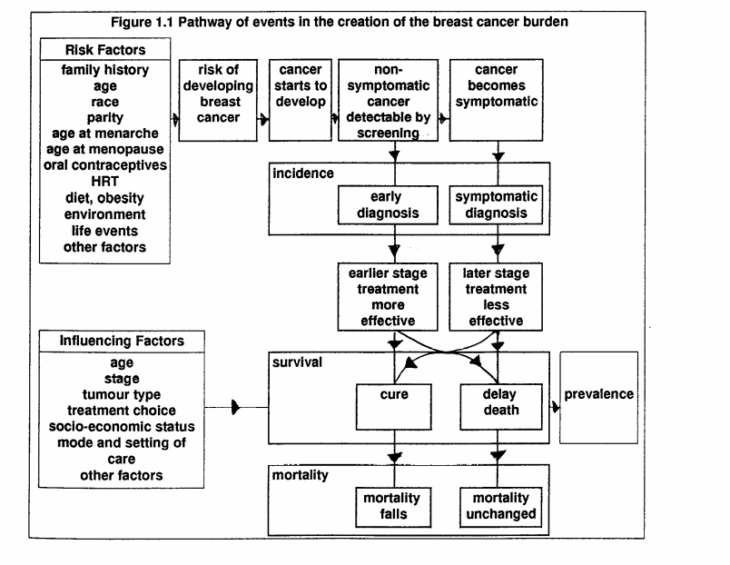
This research paper hopes to explore the pathways through breast care services in the United Kingdom. As such, risk factors like family history of breast cancer, age and parity shall be explored. In addition, the various screening processes often employed in the detection of non-symptomatic cancer shall be explored. This is important, to make an early diagnosis of breast cancer, thereby reducing the case fatalities (Familial Breast Cancer 2004).
Furthermore, the symptomatic diagnosis shall also be evaluated. About the treatment of breast cancer attending to it during the very initial stages is seen as a more effective way of handling the disease, thereby increasing the chances of cure. Conversely, treatment of the same at a later stage may prove to be less effective, with possible causation of death.
Even then, cancer treatment shall be dependent on some influencing factors that include the type of tumor, age of a patient, the choice of treatment adopted among other factors (The Breast 2007). In light of this, this research paper hopes to explore the various forms of treatment that cancer patients in the UK are usually subjected to. The various breast cancer care services experiences of cancer patients shall also be highlighted. Ultimately, this research paper shall also seek to assess the care given to the cancer patients following the various treatment methods for the disease, and the consequent fatality incidences, or lack of.
Pathway through breast cancer care services
Risk factors to breast cancer
A majority of the breast cancer cases that often affect patients in the developed countries may be explained on the basis of a number of factors that revolves around oestrogen exposure, as well as hormonal and reproductive factors, alcohol, obesity and physical activities (National Breast and Ovarian Cancer Centre (2008).
Age as a risk factor to breast cancer
Age only comes second to gender, as a strong link towards breast cancer causation. As such, older women have a higher likelihood of getting breast cancer, compared with the younger women. Well over 80 percent of the breast cancer cases in the United Kingdom have been shown to occur amongst women of above 50 years of age (Maddams et al 2008). It is this group therefore, that the National Breast Cancer Screening Programme often targets.
Statistics in the UK also shows that women between the ages of 50 and 69 are at a higher risk of being diagnosed with breast cancer, more than any other age group (Office for National Statistics 2008). The occurrence of breast cancer among teenage women or those in their early 20s may be limited, yet this is the type of cancer that is frequently diagnosed in women who are below the age of 35 years. On an annual basis, close to 1,500 women between the ages of 35 and 39 years gets diagnosed with breast cancer in the UK. The incidences of breast cancer are seen to increase with an increase in age, and the higher rate has been shown to be just before the onset of menopause, an observation that appears to reinforce the hormonal status link (Coleman 2000).
Breast cancer vs. reproductive history
When compared with their counterparts form the developing nations, women in the countries that are developed show a higher risk to cancer of the breast. This is mainly attributed to the fact that those in the developed countries tends to have less children , in relative to women in the third world countries ( The Breast 2007). In addition, the breastfeeding period of women from the developed countries is also exceedingly shorter.
According to estimates by Gotzsche et al (2009), the collective cancer of the breast incidences amongst women from the developed countries may as well be reduced to half what they are today (to 2.7, down from 6.3 persons in every 100 women), should the average births be increased to 6.5 down from 2.5 births. In addition, cancer incidences were also estimated to reduce following extended periods of breast feeding up to 24 months (Lancet 2002).
Factors of reproductive health affecting the risk to breast cancer
There is a strong association between a rise in breast cancer risk and an early menarche age. For the developed countries, the standard menarche age is today estimated at between 12 and 13 years, a drop from an average of 16 and 17 years during the middle part of the 19th century (Halperin et al 2007).The more the menarche age gets delayed, the lesser the risk factor for one to develop cancer of the breast. This is the case with such countries like China that enjoys a standard ‘age at menarche’ of 16 and 17 years (Halperin et al 2007).
There is also a correlation between the age at which a woman gets her first child, with the risk for cancer of the breast increasing with an increased age at first birth (Dixon 2006). For every one year that a woman delays in getting a child, breast cancer risk tends to increase by about 3 percent (Dixon 2006).The age at which one gets the first birth is also a factor. There is also an association between parity and the consequent reduction in cancer of the breast risk factors, with those women bearing greater number of children having a reduction in terms of the risk factors to breast cancer.
Breastfeeding has also been shown to reduce the risk factors to cancer of the breast, with a longer during of this exercise further reducing the risk factor. (Dixon 2006). Furthermore, breast cancer is seen to increase amongst those women that experience a late stage of menopause, with those women that have not attained the period of menopause enjoying a relatively lesser breast cancer risk.
Breast cancer and diet
There is an association between breast cancer and a high fat diet, at least from animal studies, and several case control studies (The Breast 2007). Nevertheless, a combination of cohort studies failed to discover a profound correlation between a risk of getting breast cancer, and the intake of a diet high in fat (Dixon 2006; The Breast 2007). On the whole, there exists evidence to support claims that the consumption of a diet high in animal fat translates into an elevated risk to cancer of the breast.
Family history
There is a double risk of a woman with a cancer case history in her family to later in life be affected by cancer herself, compared to one with no family history of breast cancer. The risk appears to increase as the number of cancer victims in a family increases (Imaginis Corporation 2006). Nevertheless, more than 85 percent of the women whose close relative(s) have been victims of cancer of the breast may never get affected themselves (Office for National Statistics 2005). A third of the cases of breast cancer amongst the developed nations are often attributed to heredity, with the remaining two thirds being attributed to lifestyle and environmental factors (Palmieri 2000).
Screening to detect non-symptomatic cancer
Breast cancer mortality rates have fallen in the UK, thanks to the efforts by the NHS, through their breasts screening programme initiative, but the disease still remains the leading cause of death amongst women in the UK. Breast cancer screening has been projected to result in an early diagnosis of the disease, thereby leading to treatment procedures likely to result in a cure, and not fatalities (Dixon 2006). Basically, screening is supposed to detect the tiny breast cancer tumours, prior to the development of metastases (The Breast 2007).
So that the breast screening exercise may be effective, the early developmental stages ought to be quite recognisable. This therefore represents the phase of preclinical detection, in the case of cancer of the breast (Dixon 2006). This is the phase at which a mammography facilitates in the detection of tumours prior to their becoming palpable. At such a stage, the tumours (often measuring a cm in diameter) have a reduced chance of turning invasive. In the event that they have already turned invasive, chances of them spreading locally or at a distance are also quite remote (Rusiecki et al 2005).
In the UK, breast cancer screening came into being as a result of the Forrest Report recommendations, whose publication was in 1986. Initially, the NHS programme for breast screening (NHSBSP) sough to provide ‘single mediolateral oblique-view mammography’ to those women whose age was between 50 and 64 years, within a 3 year interval (Dixon 2006). However, research advances in breast cancer in the UK have led to even women as old at 69 years getting invitations to join the programme, in addition to the provision of a ‘two-view mammography at all screens’ (Rusiecki et al 2005).
There is a need for repetitive breast cancer screening exercise to be undertaken on a periodic basis, so that its effectiveness may be ascertained. This is because as one ages, there is a higher likelihood that one may develop cancer of the breast. Moreover, there is a variation with regard to the disease rate of growth, as the years pass (The Breast 2007). Nevertheless, a compromise requires to be reached between on the one hand, how practical it would be to undertake periodic screening versus the associated cost, while at the same time also ensuring that the number of cancer cases that evades detection gets reduced drastically (Dixon 2006). The screening interval in the UK had previously been established at 3 years, and this is the interval that the NHSBSP has adopted to-date (NHSBSP 1991).
Modalities of screening
Three fundamental tools for breast cancer screening have been established: the use of a mammography, self examination, or having a trained medical staff examining a patient’s breasts physically (Wilson & Houssami 2006). Mammography makes use of ionizing radiation for purposes of imaging the tissues of the breast. To accomplish the exercise, the breast is often firmly compressed between a cassette of x-ray containing a special film of x-ray, and a plastic plate. In case of a routine check-up, the films are normally taken “in mediolateral oblique and craniocaudal projections” (Dixon 2006). The two views encompass the tissue of the breast up to the pectoral muscle right from the breast nipple (Maddams et al 2008).
The level to which a mammography may turn out to be sensitive varies, depending on the conspicuity and size of a lesion, the density of the breast tissue, age of the patient, the tumours’ hormonal status, the radiologists’ interpretative techniques, as well as the overall quality of the image (Dixon 2006). Mammography is capable of detecting breast cancers too minute to palpate following a physical examination, in addition to the detection of ‘ductal carcinoma in situ (DCIS) that is often characterized as a ‘noninvasive condition’ (Dixon 2006)..
A self-breast examination gets done by a patient on their own accord, a practice that in the UK is now greatly advocated for (usually, at least once every month), so as to increase the chances of early breast cancer detections, and thereby reduce the risk of breast cancer (Maddams et al 2008). Amongst the breast clinics that this researcher visited, well over 50 percent of the patients that were in attendance of the clinics confessed that they first undertook self-examination of their breast, at which point they felt unusual lumps, thereby prompting them to seek medical attention.
Still, the feeling amongst a greater majority of the patients is that the NHS, through its breast screening programme, has done a commendable job of not only creating awareness about this leading cause of deaths amongst the women in the UK (NHS 2000), but also by way of by of ensuring that the breast cancer fatalities are reduced, following an early detection.
Symptomatic breast cancer diagnosis
In the UK about 200 units of the NHS have specialised in a diagnosis of the symptomatic breast problems. In addition, a majority of the breast cancer cases have been shown to have been detected at the referral clinics for symptomatic breast problems (Dixon 2006). This researcher, through visiting various breast cancer referral clinics, discovered that on average, about 60 percent of all the recorded cases of breast cancer had been diagnosed via a presentation of the patients with symptomatic breast problems.
The referral clinics ensure that they maintain the national protocols which describe the triple test. This is a test that involves imaging (a combination of ultrasound and mammography), a clinical assessment and core biopsy (Maddams et al 2008). In the UK especially, ‘one-stop clinics’ are usually advocated for. These are the clinics in which all the needed tests necessary to arrive at a plausible breast cancer diagnosis are made. So that an early symptomatic cancer of the breast may be achieved, women usually are encouraged by way of overall methods of health promotions, to seek help in such ‘one-stop clinics’ (Office for National Statistics 2005), in case of a development in unusual breast alterations.
Before one is referred to a ‘symptomatic breast clinic’ by a General Practitioner, there are a number of symptoms that a doctor may have come across to warrant such a decision. These may include the existence of an unusual lump at the breast, or even as a result of such a different symptom was a discharging breast or feelings of discomfort (Rusiecki et al 2005). Once this is established, the next stop in the x-ray department located in a breast clinic. At this department, either a breast ultrasound exercise, or a mammogram may be undertaken, sometimes, both these practices are undertaken. It is also not uncommon to have a biopsy test carried out at these breast clinics (Dixon 2006).
Following a successful accomplishment of the triple assessment preferably carried out by a professional; oncologist, a patient may then have their breasts imaged, usually by the use of ultrasound or a mammogram, the determining factor here being the age of a patient (Lacroix 2006). In a majority of the cases, an ultrasound tests alone shall suffice for women below the age of 35 years. This is important, so that the minor risk that accompanies an x-ray may be avoided.
In addition, the breast tissue of younger women tends to be extremely dense, wit the result that changes of the breast may not feature on an x-ray. Ultimately, cells or fluids could be removed from a lump in the breast by way of using a tiny needle (Lacroix 2006). This is a technique of ten referred to as ‘fine needle aspiration’ (FNA). Occasionally, it may become necessary that a lump’s ‘core biopsy’ is performed. This involves the removal of a minute piece of the breast lump via the use of a somewhat bigger needle (Dixon 2006).
Breast cancer survival influencing factors
Following a successful diagnosis on the breast cancer through non-symptomatic screening, there is every possibility of a patient benefiting from a timely and effective treatment and a possible cure, with the result that the mortalities of breast cancer falls (Imaginis Corporation 2006).
On the other hand, if one were to wait for a symptomatic diagnosis, what this means that the diagnosis shall have been delayed, a delay that could result in the death of the patients, in effect translating into higher rate of prevalence of breast cancer-related deaths (Imaginis Corporation 2006). Nevertheless, there are a number of influencing factors with regard to the survival of breast cancer patients.
The age of the patient is very important, with the survival rates shown to decrease with an increase in age (Dixon 2006). This is because of the successive treatment regimens that may accompany a diagnosis of breast cancer, such as the use of chemotherapy, radiotherapy or hormonal replacement therapy. As such, the tolerance levels of the elderly may be lower compared to say, a young woman. The stage at which the breast cancer gets diagnosed is also another factor (Maddams et al 2008). Not only are the late stage cancer tumours bigger, they also tend to invade extensively, and this acts to reduce the survival rate of the breast cancer victims.
The type of tumour is yet another factor, with the invasive types being more difficult to manage in comparison to the non-invasive (Gotzsche et al 2009). Furthermore, the choice of treatment shall also take a tool on the health of an individual. Another factor worth considering is the socio-economic status of the breast cancer victim. When a patient is financially well-off, chances are that they are also in a position to manage the disease more effectively, in comparison to those patients that are not financially-well off. The mode of care that the breast cancer patient receives shall also determine whether or not they have a chance of surviving (Gotzsche et al 2009).
Treatment of breast cancer patients
Regardless of the method of diagnosis involved (whether it is the symptomatic screening or the symptomatic assessment), once a patient is diagnosed with breast cancer, the treatment methods adopted are more or less similar. Nevertheless, each breast cancer patient is handled as an isolated case, different from any other patient. Generally, the kind of treatment that a breast cancer patient receives will be dependent on several factors such as the age of a patient, the stage as well as the grade of the cancer, if at all a woman has attained the menopause stage, tumour size, and whether or not the cells of the cancer bears receptors for specific hormones (for example, oestrogen) or even certain proteins, like HER2 (Rusiecki et al 2005).
A majority of the breast cancers shall often get treated by way of surgery, with a view to removing the tumour. In this case, all, if not part of the breasts, may require to be surgically removed. In a case whereby the entire breast is surgically removed, it might become necessary for such a patient to undergo a breasts reconstruction (Rusiecki et al 2005). This procedure could run concurrently with the surgery. It could even have taken place prior to the surgery, or come in later.
From time to time, it may be necessary to have breast cancer patients undergo either a hormonal therapy or a chemotherapy session, with the aim of assisting in the shrinking of the cancerous cells, prior to the actual surgical removal of these cells. It is this technique that has come to be known as ‘neo-adjuvant therapy’. Immediately upon surgery any remaining tissue of a breast may be subjected to radiotherapy (Familial Breast Cancer 2004).
In a case whereby a breast has had to be removed (mastectomy), such radiotherapy may have to be given to the wall of the chest. The reason for undertaking this radiotherapy procedure is to ensure the destruction of cancerous cells that could have escaped the surgical removal. It is only after surgery that the doctors are in a position to comment on both the grade and stage of the cancer. In addition, it now becomes possible to forecast on the likelihood of the cancer either re-occurring or spreading later (The Breast 2007).
Some of the factors that could result in a re-occurrence of cancerous cells includes the tumour size, if at all the lymph nodes located in the armpit got affected, the tumour gland, if at all the cells of the cancer have invaded the blood or lymph vessels that bordered the tumour, if at all the cancerous cells bore on their surface oestrogen receptors, or such specific proteins as HER2 (Gotzsche et al 2009). In the short-term, the likelihood of cancerous cells with receptors for oestrogen recurring is quite remote, whereas those bearing HER2 receptors are almost guaranteed of recurring, only if the patient receives Herceptin.
For a patient whose chances of cancer spreading are quite remote, there is no dire need for them to undergo further treatment (Lacroix 2006). Nevertheless, in the event that the cancerous cells might come back, a majority of the women, especially those in whom the oestrogen receptor is negative, these are often advised to undergo a chemotherapy treatment. On the other hand, for the patients whose oestrogen receptors are positive, these are often advised to undergo a hormonal therapy. (Both of these constitute the adjuvant therapy).
Recovery and rehabilitation of cancer survivors
Given the fact that cancer as a disease is very much draining in terms of emotional and physical energy, there is a need therefore to ensure that once the patient have received the necessary curative or palliative treatment, that they are then placed in a recovery and rehabilitation centre in which they shall make a progressive recovery under the watch of medical assistance (Buchsel & Yarbro 2005). In such recovery and rehabilitation centres, this is where the patients are in a position to receive educational support as regards the cause, the risk factors, symptoms, treatment, and management of breast cancer.
Furthermore, the patients stands a chance to gain the knowledge of whether or not they are predisposed to experience a recurrence of the cancer, based on the size of the tumours, and the grade of the cancer, among other factors. In addition, this educational support could be provided on an individual basis, or to a group of cancer patients (Rusiecki et al 2005). Moreover, the implementation of a cancer rehabilitation program may also entail training to the patients of the lifestyle and nutrition behavioural changes that they may be expected to embrace, following the development on their medical history.
This is more of a counselling program, seeing that there are habits that a patient may have to do away with, and at the same time embrace alien habits. As such, a psychological counselling beforehand is often deemed necessary so that a patient may not slump into a stress-induced mode, thereby hampering any recovery efforts.
Besides, there is a need to have in place partner or peers to the patients visiting them at the recovery and rehabilitation centres. All of these could as well be incorporated in the education and counselling program. This is because the peers and the partners are the ones who are more acquainted with the patient, and as such, they are at a better position to alter the behaviours and patterns of their loved ones who are sick (Halperin et al 2007).
Furthermore, the recovery and the rehabilitation programs should also take into account such elements as the prosthetics, boutiques, scarves and wigs. These are a necessity for the cancer patients, as one of the side effects of the therapy sessions especially chemotherapy is a loss of hair (Dixon 2006). For this reason, there is a need to ensure that the patient do not perceive as if they have lost their self-esteem for example, along with the lost hair, and so the more reason why these facilities ought to be incorporated in the recovery and rehabilitation program.
Conclusion
Breast cancer has emerged as the most common type of cancer amongst women in the United Kingdom, especially amongst the women past the age of 50 years with the occurrence rate projected at 8 out of every 10 women (Office for National Statistics 2008). Perhaps as a cue to this, the NHS runs a breast cancer diagnostic programme that targets women between the age of 49 and 53 years, although it has lately been modified to accommodate those as old as 69 years. This is aimed at ensuring that a timely diagnosis is made so that the treatment may be effective.
In the UK, a pathway through the disease burden that is breast cancer often starts with a non-symptomatic diagnosis via screening. Screening could be through the use of a mammography, self-examination by patients, or clinical examination. In case of delays, then breast cancer could also be detected via the use of symptomatic screening (Gotzsche et al 2009).
Nevertheless at this time, the cancer shall often have spread and the treatment may not be as effective, resulting in an increase in case fatalities and a resultant increased prevalence. Early diagnoses shall also determine the survival rate of the breast cancer patients, although such other factors as age, treatment choice, and the type of tumour should also be considered.
Bibliography
Advisory Committee on Breast Cancer Screening (2006). “Screening for Breast Cancer in England: Past and Future” Executive Summary Report
American College of Radiology. “Safety: Radiation Exposure in X-ray Examinations”. Radiology Society of North America. (2006). Web.
Cancerbackup (2009). “Risk factors and causes of breast cancer”. Web.
Coleman, M. “Trends in breast cancer incidence, survival, and mortality. Lancet, Vol. 356, No. 9229 (2000): 590.
Dixon, M. J, 2006, Breast surgery. London: Elsevier Health Sciences.
Familial Breast Cancer. “The Classification and care of women at risk of familial breast cancer in primary, secondary and tertiary care”. National Institute for Clinical Excellence, 2004.
Finkel, M. L. (2005) “Breast cancer and treatment options” Understanding the mammography controversy pp 39.
Gotzsche, P. C, Ole J Hartling, O. J, Nielsen, M, Bodersen, J & Jorgensen, K. J. “Breast screening: the facts-or maybe not” BMJ 2009 338:b86
Halperin, E, Perez, C, & David, E, 2007, Perez and Brady’s principles and practices of radiation oncology. Amsterdam: Wolters Kluwer Health.
Imaginis Corporation.” Breast cancer: statistics on incidence, survival, and screening”. Imaginis Corporation, 2006. Web.
Lacroix, M. “Significance, detection and markers of disseminated breast cancer cells”. Endocr Relat cancer Vol. 13, No.4 (2006): 1033–67.
Maddams J, Moller, H & Devane, C. “Cancer prevalence in the UK, 2008. Thames Cancer Registry and Macmillan Cancer Support.
NHSBSP, 1991, “Breast Cancer Screening 1991: Evidence and Experience since the Forrest Report”.
National Breast and Ovarian Cancer Centre (2008). Breast symptoms and diagnosis. Web.
NHS “Effect of NHS Breast Cancer Screening Programme on Mortality from Breast Cancer in England and Wales, 1990-8: Comparison of Observed with Predicted Mortality”. BMJ 2000:665-669).
Office for National Statistics, Cancer statistics registrations: registrations of cancer diagnosed in 2005, England. Series MBI No. 36. 2008, National Statistics: London.
Palmieri, C. “Breast cancer screening has to be combined with good surgical and oncological services” BMJ. Vol.321, No.7260 (2000): 567.
Rusiecki, J. A, Holford, T. R, Zahm, S. H & Zheng, T (2005). “Breast cancer risk factors according to joint oestrogen receptor and progesterone receptor status”. Cancer Detect. Prev. Vol. 29, No. 5 (2005): 419–26.
The Breast. “Metastatic breast cancer: recommendations proposal from the European school of oncology (ESO) – MBC task force. The breast, Vol. 16 (2007): 9-10. Web.
Wilson, R, & Houssami, N “Should women at high risk of breast cancer have screening magnetic resonance imaging (MRI)?” The Breast (2006): 2-4. Web.
Wilson, R, & Liston, J. “Clinical guidelines for breast cancer screening assessment”. NHS Cancer Screening Programmes. Vol 49 (2005): 3-11.
Yager, J. D, & Davidson, N. E. “Oestrogen carcinogenesis in breast cancer “. New Engl J Med Vol. 354, No. 3 (2006): 270–82.
Appendices