Introduction
The heart is one of the essential organs in one’s body. It is responsible for moving nutrients and oxygen to the tissues and organs, acting as a pump, and transferring venous and arterial blood in several directions. This organ keeps the body satiated with the vital elements for its existence and healthy performance. The veins and arteries dispersed through the limbs and organs connect to the heart, and this central organ located in the center of the rib cage is linked to every part of one’s body. The pressure inside the heart and this organ’s processes determine the cells’ oxygen saturation.
Labeling the Heart: Structure and Function
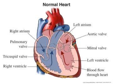
As demonstrated in Figure 1, the heart can be visually separated into four main chambers. First, the two upper segments are atria – they collect blood that moves into the heart (National Heart, Lung, and Blood Institute [NHLBI], 2022). Below them, one can find two ventricles – they pump the collected blood out of the heart and into other organs and tissues of the organism (NHLBI, 2022). The connecting tissue between these areas is called a valve – this opening manages blood flow and stops it from flowing in the wrong direction. The heart is also separated vertically, creating a left and right side with one ventricle and atrium.
Blood Flow Through the Heart, Lungs, and Body
The cycle of moving blood can be separated into venous and arterial flow. To begin with, blood deprived of oxygen that has traveled through the entire body returns to the heart through the veins – superior and inferior vena cava (NHLBI, 2022). It enters the right atrium and moves to the right ventricle through the tricuspid valve. Then, exiting the right ventricle and passing the pulmonary valve, the venous blood goes directly to the lungs, which fill it with oxygen (NHLBI, 2022). As an outcome, venous blood becomes arterial and becomes ready to deliver the necessary oxygen to body tissues.
Blood filled with oxygen returns to the heart’s other part – it enters the left atrium via the pulmonary veins that connect the lungs to the heart (NHLBI, 2022). It moves through the mitral valve into the left ventricle, which pumps the blood into the aorta and the rest of the body through the aortic valve (NHLBI, 2022). The aorta separates into arterioles and much smaller channels called capillaries, which bring oxygen-rich blood to the organism’s cells. There, as organs take the oxygen and capillary blood transfers it into venules, it transforms into venous blood and moves towards the heart.
This process shows that the heart acts as a pump, receiving and pushing blood to reach every part of one’s body. Thus, the heart must operate under constant pressure and maintain balance in its segments to transport liquids across the entire system effectively.
Normal Pressures in Each Segment of the Heart and Vessels
Standard pressure measures are established in each element and vessel that carries the blood. The average proper atrium pressure (RAP) is 3 mm Hg, although it may shift from 0 to 8 mm Hg throughout the cycle (MSD Manual, 2023). The pressure in the right ventricle (RVP) can be estimated at 25 mm Hg (15-30 mm Hg) during systolic and 4 mm Hg (0-8 mm Hg) at diastolic phases (MSD Manual, 2023). Central venous pressure (CVP) defines the pressure in the vena cava and is around 2 to 6 mm Hg (MSD Manual, 2023). These averages represent the blood circulation in the right part of the heart.
In the left segment of this organ, the pressure is especially vital as it is linked to how the blood will move out of the heart. Pulmonary artery pressure (PAP) is mean at 15 mm Hg (9-16 mm Hg), and its systolic and diastolic measures are 25 mm Hg (15-30 mm Hg) and 9 mm Hg (4-14 mm Hg), respectively (MSD Manual, 2023). The pressure in the pulmonary artery wedge (PAWP) is around 9 mm Hg (2-12 mm Hg) (MSD Manual, 2023).
In the left ventricle, the pressure is incredibly high – typically, it is between 90 and 140 mm Hg during systolic phases and 5 and 12 mm Hg during diastolic phases (MSD Manual, 2023). The two phases in the left atrium are A and W waves, and their pressure is about 4-16 and 6-12 mm Hg (MSD Manual, 2023). These measures constantly fluctuate as the heart pumps the blood, but the range stays within these standard values if the organ can maintain balance.
As noted above, the pressure inside the heart and its vessels determines how other organs will be supplied with oxygen. The concept of cardiac output is used to measure how much blood exits the left ventricle. It is the volume of blood pumped out of the heart each minute, and it is calculated by multiplying the heart rate by the stroke volume (Pollock & Makaryus, 2022).
In the end, one gets a number that describes how many liters of blood exit the heart each minute. To determine whether the output is sufficient, comparing it to the person’s age, weight, and other characteristics is essential. Here, stroke volume. A decrease may imply that the heart cannot pump much blood – a low heart rate and decreased pressure reduce the values, while increased preload and a good tone may increase the output.
Preload, Afterload, Cardiac Output, and Systemic Vascular Resistance (SVR)
Next, SVR refers to systemic vascular resistance – the amount of force the body needs to circulate the blood around. It depends on blood viscosity and arterial tone, as thinner blood requires less power, while more viscous blood can increase the values (Trammel & Sapra, 2023).
The concepts of preload and afterload are crucial for determining the blood volume in the heart. Preload describes how much blood is already present in the ventricles, and afterload is the pressure needed to pump the blood out of the left ventricle into the system. Increased preload and afterload may be caused by tissue degradation, heart failure, and high blood pressure (Pollock & Makaryus, 2022). At the same time, analgesics may relax the heart and decrease these values (Pollock & Makaryus, 2022). The balance between the two ensures that the blood will quickly exit the heart and the demand and supply will be maintained.
The Importance of Proper Circulation for Tissues and Organs
The heart provides tissues and organs with oxygenated blood and circulates the venous blood to the lungs, maintaining the cycle. Proper circulation ensures that all cells have the nutrients necessary for survival and functioning. As an outcome, one’s body performs as well as it can, having an active immune system, cell regeneration, healing, and energy (NHLBI, 2022). Blood circulation is also responsible for temperature regulation and healthy skin, nails, and hair.
Conclusion
In conclusion, the heart is responsible for supporting the whole body, and its performance determines the activity of other organs. Oxygen circulation revolves around the heart, which is the central pump for arterial and venous blood. The blood moves through the right and left sides of the heart, going from and to organs with the necessary elements. The different pressures inside the heart and vessels determine how much force is needed to pump blood, and various factors impact the organ’s effectiveness.
References
MSD Manual. (2023). Normal pressures in the heart and great vessels. Web.
National Heart, Lung, and Blood Institute (2022). What the heart looks like. Web.
Nucleus Medical Media. (2022). Normal heart [Illustration]. Web.
Pollock, J. D., & Makaryus, A. N. (2022). Physiology, cardiac cycle. StatPearls. Web.
Trammel, J. E., & Sapra, A. (2023). Physiology, systemic vascular resistance. StatPearls. Web.