Definition
Ebola Virus Disease (EVD) is an acute natural focal zoonotic viral infection characterized by febrile intoxication syndrome, phenomena of universal capillary toxicosis with a severe hemorrhagic syndrome, multiple organ lesions, and a high mortality rate. According to the antigenic properties of glycoproteins (GP), there are five types: Bundibugyo ebolavirus (BDBV), Reston ebolavirus (RESTV), Sudan ebolavirus (SUDV), Tai Forest ebolavirus (TAFV), and Zaire ebolavirus (EBOV) (Hu et al., 2017). Four of them cause diseases of different severity in people in Africa (TAFV – lethality 0%, BDBV – lethality up to 30%, SUDV – lethality up to 50%, EBOV – lethality up to 90%) (Caviness et al., 2017). Zaire ebolavirus is considered a type species of the genus. Manifest cases of RESTV disease, highly pathogenic for monkeys, have not been identified in humans.

People become infected with the Ebola virus through either contact with infected animals (usually after slaughtering, cooking, or eating) or contact with body fluids from infected people. Most cases are caused by human-to-human transmission, which occurs when blood, other fluids, or secretions (feces, urine, saliva, semen) from an infected person enter a healthy person through damaged skin or mucous membranes. Infection can also occur when damaged skin or mucous membranes of a healthy person come into contact with objects or the environment contaminated with an infected person’s body fluids. These can include soiled clothing, bedding, gloves, protective equipment, and medical waste, such as used hypodermic syringes.
Classification
- Class: Monjiviricetes
- Order: Mononegavirales: RNA viruses with contiguous genomes
- Family: Filoviridae
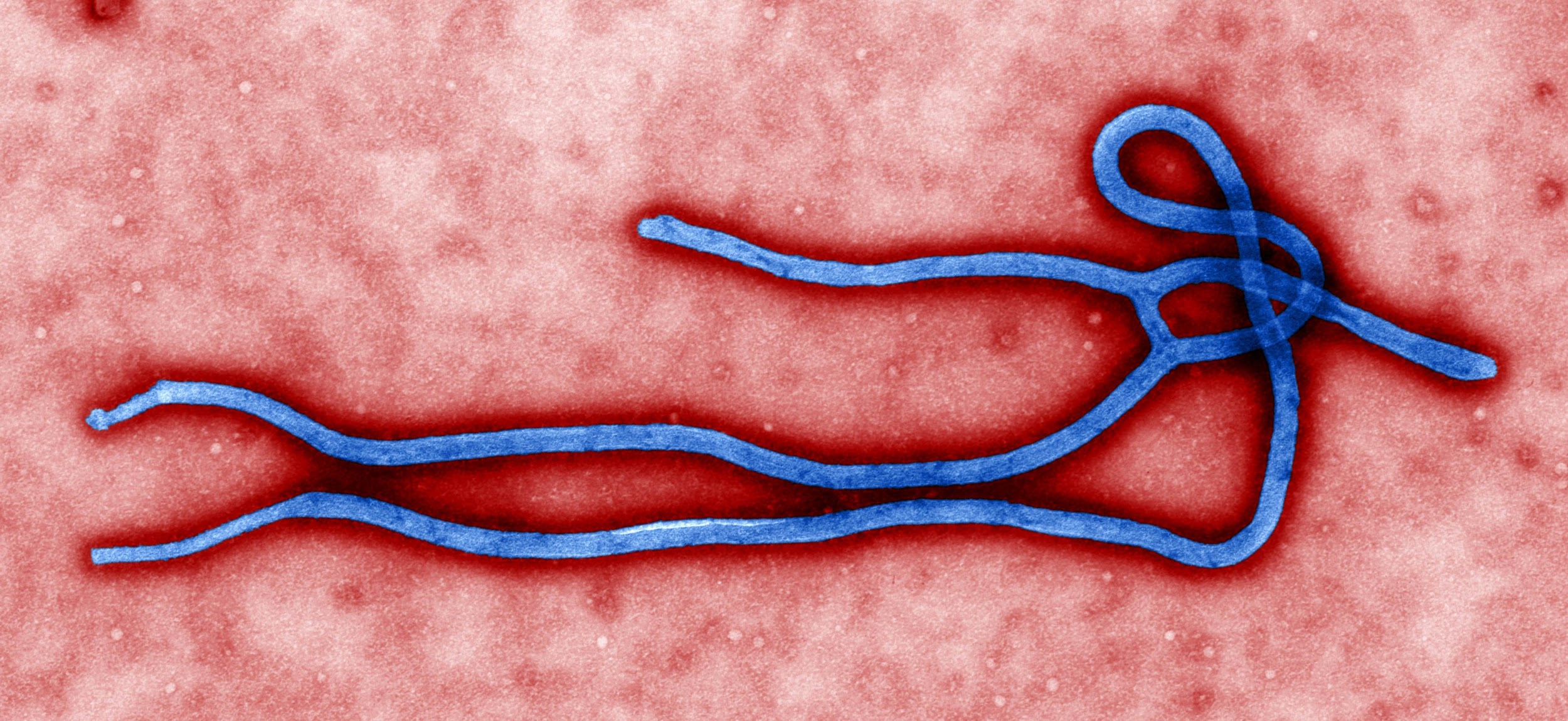
The Ebola virus belongs to the filoviridae family. The family name comes from lat. Filum, a filament, and reflects the morphology of virions. The family includes two genera: Marburgvirus and Ebolavirus. Viruses of this family cause severe hemorrhagic fevers, often fatal. The filoviridae virion has a lipid envelope and the form of twisted filaments 600–800 nm long and 50 nm thick. The nucleocapsid is a complex of viral RNA and four structural proteins: NP (nucleoprotein), VP30 (viral polymerase co-factor), VP35 (phosphoprotein), and L (viral RNA-dependent RNA polymerase) (Kuhn et al., 2019). Three structural proteins are associated with the virus membrane. Two of them, VP24 and VP40, are located on the inner side of the membrane and play the role of matrix proteins, while the surface GP-complex, consisting of two subunits (GP1 and GP2), forms the outer spines of the virion (Kuhn et al., 2019). The filoviridae genome is a single-stranded RNA of negative polarity, about 19,000 base pairs in size, flanked by conserved non-coding regions. The genome contains seven open reading frames – one for each gene.
- Species: Ebolavirus
The virion has the form of long filaments, variously twisted (V-shapes, the shape of the number 6 or spiral shapes), 70-100 nm in diameter, and 974-1086 nm in length. The nucleocapsid consists of a central axial filament with a diameter of 40-50 nm. The genome is represented by a single-stranded negative RNA (18959-18961 nucleotides) surrounded by a lipoprotein membrane with spikes 7-10 nm long. The virus contains seven structural proteins (NP, VP35, VP40, GP, VP30, VP24, L) (Zinzula et al., 2019). Viruses from the genus Ebolavirus are similar in morphology to Marburg marburgvirus but differ in antigenic structure. The viruses are highly variable: under laboratory conditions, cultures of the pathogen are maintained by the passage in cell culture of guinea pigs and Vero with a mild cytopathic effect (Caviness et al., 2017). Viruses have an average level of resistance to damaging environmental factors. The virus persists at -20 ° C throughout the year (Caviness et al., 2017). At 4 ° C (in the refrigerator when storing donor blood), viruses’ pathogenic properties do not decrease within five months. Viruses are resistant to ultraviolet radiation. They are reliably inactivated by formalin and 0.5% chloramine B when mixed with culture 1: 1.
References
Caviness, K., Kuhn, J. H., & Palacios, G. (2017). Ebola virus persistence as a new focus in clinical research. Current opinion in virology, 23, 43-48. Web.
Hu, J., Jiang, Y. Z., Wu, L. L., Wu, Z., Bi, Y., Wong, G.,… & Zhang, Z. L. (2017). Dual-signal readout nanospheres for rapid point-of-care detection of ebola virus glycoprotein. Analytical chemistry, 89(24), 13105-13111. Web.
Kuhn, J. H., Amarasinghe, G. K., Basler, C. F., Bavari, S., Bukreyev, A., Chandran, K.,… & Griffiths, A. (2019). ICTV virus taxonomy profile: Filoviridae. The Journal of general virology, 100(6), 911-916. hWeb.
Zinzula, L., Nagy, I., Orsini, M., Weyher-Stingl, E., Bracher, A., & Baumeister, W. (2019). Structures of ebola and reston virus VP35 oligomerization domains and comparative biophysical characterization in all ebolavirus species. Structure, 27(1), 39-54. Web.