Abstract
Fluoroscopy is a crucial diagnostic imaging modality used in medical procedures to provide real-time images of internal structures. Unlike standard radiography, fluoroscopy involves continuous or prolonged X-ray exposure, requiring specialized safety measures. The main elements of a fluoroscopy system are an X-ray tube, collimator, and image intensifier. A typical fluoroscopic procedure lasts 4 to 5 minutes.
As a result, fluoroscopic processes pose adverse radiation risks to both patients and operators. However, the use of pulsed fluoroscopy can help protect patients from radiation, while protective shields and collimators can help protect operators from stray radiation. Safety Code 35 provides guidelines on the installation, operation, and safety measures for the use of fluoroscopic apparatus.
Introduction
This report analyzes fluoroscopy imaging, highlighting its principles, components, and development. The scope of this research is limited to the technical aspects of digital fluoroscopic systems. These include the design, operation, and components of the equipment. The audience targeted includes healthcare professionals, radiologic technologists, medical students, and researchers.
Regarding background, a major milestone has been reached in medical imaging: fluoroscopy now provides video transmission. This advancement requires knowing its implications and future benefits. The report explores digital fluoroscopy, discussing its significance in real-time imaging, functional blocks, and safety issues.
Discussion
Theory
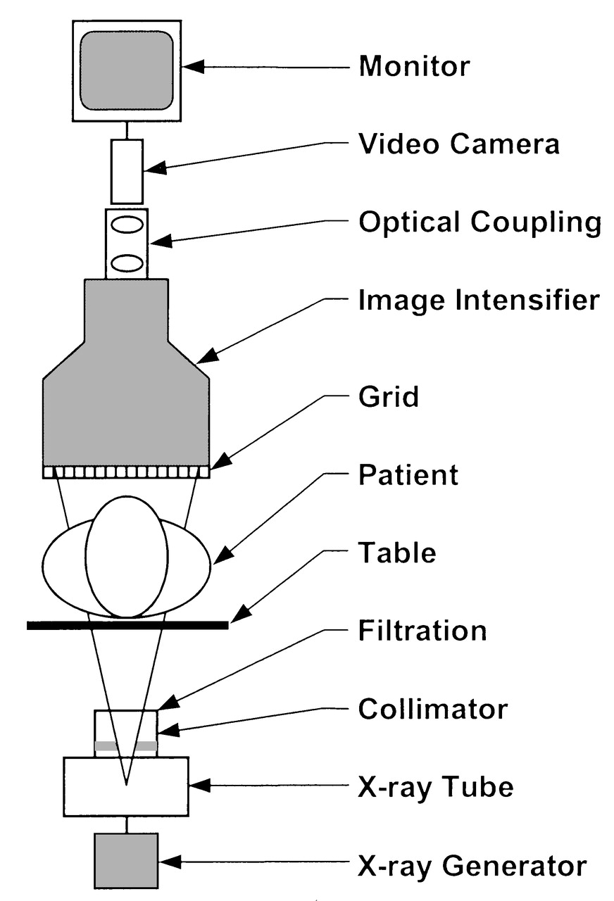
Fluoroscopy produces real-time images of a patient’s internal organs and tissues. Unlike conventional projection radiography, which captures a fixed image on film, this technology provides continuous visualization [1]. As noted in [2], some components of a fluoroscopy system include an X-ray tube that emits X-ray photons used to create images, and a collimator that shapes and limits the radiation spillover outside the required area [2]. Additionally, the image receptor (XRII) captures X-ray photons and converts them into digital images. The image intensifier (IIT) amplifies the X-ray signal before it is transmitted to the XRII, thus enhancing image clarity and minimizing radiation exposure.
Patient safety, such as preventing radiation skin injury, is required during the procedure. In digital fluoroscopy, the IIT found in conventional systems is substituted by a flat-panel detector (FPD). Additionally, the former provides static or spot pictures while the latter gives live video images [10].
The FPD converts X-ray photons into digital signals for immediate visualization. Digital detectors offer advantages, including faster turnaround times, clearer image quality, and lower radiation dose. Still, they are also associated with relatively high radiation doses and extended learning curves for radiologists, depending on the procedure [3], [4], [5].
High frame rates are required to maintain continuous motion perception. In this case, 30 frames per second (fps) is achieved by keeping the radiation dose per frame to a low of about 0.1% [2]. This produces video-like images without compromising patients’ safety. A recent development in fluoroscopy is the introduction of an AI-based eye-tracking system to improve focus, thereby reducing unnecessary radiation exposure for patients and operators during fluoroscopic endoscopy procedures [6]. This is expected to improve diagnostic accuracy and efficiency in clinical practice.
Applications
Fluoroscopy imaging is widely used in various medical procedures, including the barium swallow. This is a special imaging procedure that uses barium and X-rays to produce images of the upper gastrointestinal (GI) tract, including the throat, pharynx, and esophagus [1],[2],[4],[7]. Fluoroscopy can screen various conditions, including ulcers, hiatal hernia, and gastroesophageal reflux disease (GERD) [1]. In this case, a barium swallow enables real-time visualization of the digestive system and aids in diagnosis and treatment planning.
From a technical point of view, a fluoroscopy machine emits short pulses of X-ray beams. This precedes a patient taking a chalky liquid containing barium, a contrast agent that enhances the visibility of X-ray images [8]. X-ray radiation passes through the upper GI tract, creating real-time images displayed on a computer screen or monitor. The kV (kilovoltage) and mA (milliamperage) values for fluoroscopy are set between 80 kV and 120 kV and 100 mA to 500 mA to facilitate the required balance between patient safety and image quality [9]. Higher ranges are needed for better tissue penetration and visualization.
From the patient’s point of view, the clinic process starts by removing clothing or other objects that may interfere with the swallowing test, then lying on their back. The procedure length, based on FDA standards, is typically 4.5-5 minutes [7]. The aim is to achieve high-quality diagnostic imaging in a safe, time-appropriate manner, with patient safety as a priority. Individuals being diagnosed are required to take the barium solution beforehand so that the fluoroscopy machine can obtain real-time X-ray images of the esophagus and stomach [1]. The radiologist closely monitors fluoroscopic images to identify any deviations in the GI tract.
Description and Graphics
A digital fluoroscopic system has several functional blocks that work together to process and display fluoroscopic images. It consists of an X-ray unit, a collimator, an anti-scatter grid, an XRII, such as an IIT or FPD, an image processing unit, a display monitor, and a control interface. An X-ray source emits X-rays that penetrate the patient’s body and interact with the XRII, which converts X-ray photons to a digital image (see Figure 1).
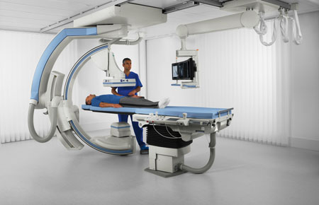
Adjusting the collimator blades maintains a consistent field of view as the source-to-image distance changes, thereby reducing radiation outside the visible area. Conversely, the anti-scatter grid is positioned between the patient and the FPD to reduce scattered radiation and improve image clarity. The signal is then passed via the image processing unit, where the adjustments to parameters, including contrast, kV, and mA, are made, and the final image is displayed on a high-resolution monitor (see Figure 2).
Safety
The risks associated with fluoroscopy procedures exceed those of conventional projection radiography. This is because the former requires continuous or serial X-rays during the imaging process, which increases radiation exposure [4]. Conversely, radiography involves a momentary exposure to X-ray radiation to obtain a single static image [10]. Since fluoroscopy provides real-time imaging, it requires long-term exposure, increasing the risk of higher radiation doses. Alternatively, in radiography, the X-ray tube is activated for only a limited time, reducing radiation-related risks.
Several features can protect patients and operators from high radiation exposure. In this case, pulsed fluoroscopy and the last-image-hold technique can reduce patient radiation exposure by lowering fluoroscopy time and overall dose [7]. Conversely, operator exposure can be reduced by using collimators and protective barriers that shield against scattered radiation [7]. Within Health Canada, the Consumer and Clinical Radiation Protection Bureau (CCRPB) is the most relevant agency responsible for regulating X-ray fluoroscopy equipment [11].
Safety Code 35 (SC35) is the main authoritative reference for Biomeds handling diagnostic X-ray tools in Canadian hospitals. The additional standards regarding fluoroscopy include the Radiation Emitting Devices Act and Regulations, which offer directives on the sale, importation, or lease of devices that produce radiation [11]. Similarly, the Canadian Nuclear Safety Commission outlines radiation safety and regulatory measures that may apply to Canadian hospitals that house fluoroscopy devices. Equally, there are territorial and provincial protocols regarding radiation safety in care facilities, including unique requirements for the usage of fluoroscopy tools [11].
SC35 contains critical instructions on the safe use of all radiation-emitting devices. The most important sections within SC35 related to medical X-ray fluoroscopy tools are A and B [12]. The former outlines the duties of personnel operating fluoroscopy equipment, while the latter offers guidelines on its effective installation and operation [12].
According to SC35, the Non-invasive X-ray tube voltage meter is the annual quality control test equipment required only for fluoroscopy tests [12]. The qualification requirements for Biomeds tasked with preserving fluoroscopic systems include knowledge and training in the repair and maintenance of radiology-related imaging equipment, as well as radiation protection guidelines and procedures.
Conclusion
The discussion has provided principles and components of the fluoroscopic system, shown how it differs from conventional radiography, and addressed related safety concerns. However, regulation by the CCRPB and SC35 can guarantee the safe and effective use of this equipment. A deep understanding of the intricacies of fluoroscopy technology will enable healthcare professionals to mitigate radiation risks without compromising diagnostic outcomes. Additionally, in the future, integrating fluoroscopy technology with AI is anticipated to increase precision in patient examinations and ensure patient safety.
References
- “Fluoroscopy Procedure.” Johns Hopkins Medicine.
- G. Eric and J. Thomas. “Modern Fluoroscopy Imaging Systems.” American College of Radiology.
- “Digital radiography and fluoroscopy.” St. Anthony Community Hospital.
- “Fluoroscopy.” Food and Drugs Administration.
- “Conventional and digital fluoroscopy.” International Atomic Energy Agency. Human Health Campus – Conventional and digital fluoroscopy (iaea.org).
- T. Masaki et al., “Eye-tracking fluoroscopy system: A new artificial intelligence-based system to communicate activate watching of the monitor during endoscopy,” Endoscopy, vol. 55, no. 1, pp. 179–180, 2023.
- P. Shellie. “Technical principles for diagnostic fluoroscopic procedures. American College of Radiology.
- V. Daniel and M. Junaid, Safety of Fluoroscopy in Patient, Operator, and Technician. Treasure Island, FL: StatPearls, 2023.
- N. Miyuki et al., “Radiation exposure and protection in computed tomography fluoroscopy,” Interventional Radiology, vol. 7, no. 2, pp. 49-53, 2022.
- “Finding the difference between an X-ray and a fluoroscopy.” Canadian Diagnostic Network.
- “Radiation-related regulations in Canada.” Radiation Safety Institute of Canada.
- “Safety Code 35: Safety procedures for the installation, use and control of x-ray equipment in large medical radiological facilities.” Government of Canada.