Introduction
The skeleton is a key component of the human musculoskeletal system. Its structure is similar to that of all mammals, but it has features associated with upright walking. The body of any person relies on the skeleton, which provides support from within. Each bone differs from the others depending on exactly where in the body it is located and what work it has to do.
Structure of the System
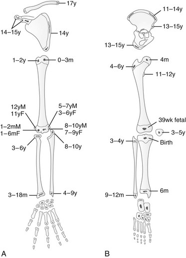
Throughout life, the skeleton undergoes constant changes. During intrauterine development, the cartilaginous skeleton of the fetus is gradually replaced by bone. This process also continues for several years after birth.
A newborn baby has almost 300 bones in its skeleton, much more than an adult (Kan & Strouse, 2019). This difference arose because the children’s skeleton contains many small bones, which fuse into large ones only at a certain age (Kan & Strouse, 2019). Figure 1 shows ossifications of the upper (A) and lower (B) limbs.
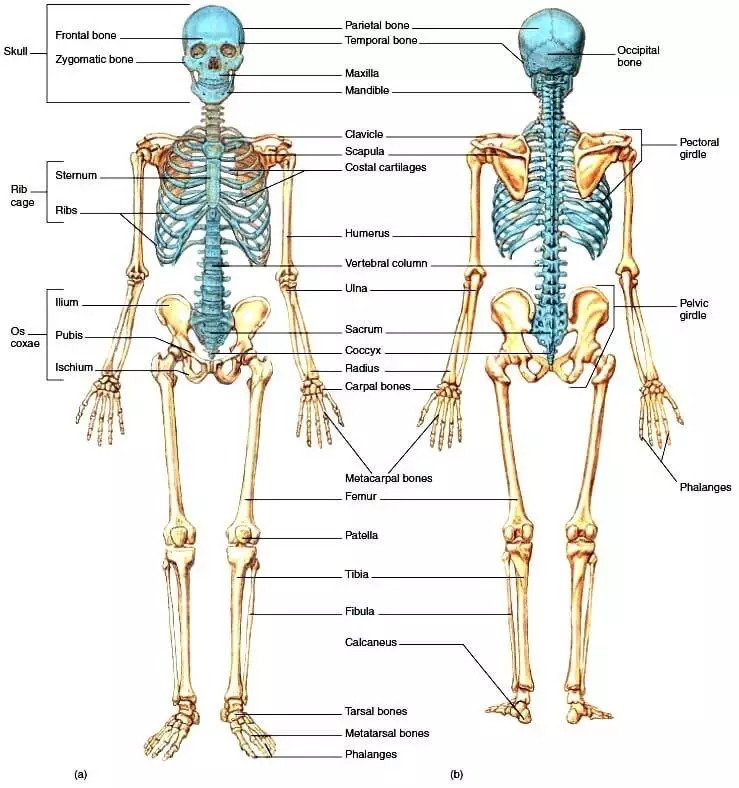
The passive part of the human musculoskeletal system is a complex of bones and their joints. The skeleton consists of the bones of the skull, spine, and chest (axial skeleton), as well as the bones of the upper limbs and lower limbs (appendicular skeleton) (Dunn, 2022). Figure 2 shows the skeletal system of an adult, its main sections, and its structure. The system is characterized by high strength and flexibility, which is provided by the way the bones are connected.
The movable connections of most bones give the skeleton the necessary flexibility and provide freedom of movement. In addition to fibrous and cartilaginous continuous joints (they mainly connect the bones of the skull), there are several less rigid bone joints in the skeleton (Dunn, 2022). Each type of connection depends on the required degree of mobility and the type of load on a given part of the skeleton. Joints that allow only limited movement are called semi-joints or symphyses, while joints that are freely movable and separated by a cavity (synovial joints) are called joints. The complex geometry of the articular surfaces corresponds to the degree of freedom of this connection.
Six particular bones (three on each side) in the middle ear do not belong directly to the skeleton. The auditory ossicles are connected only to each other and participate in the work of the organ of hearing, transmitting vibrations from the eardrum to the inner ear. Joints with limited mobility are called semi-joints or symphyses, and discontinuous (synovial) joints are called joints. The hyoid bone, the only bone not directly connected to others, is located in the neck but is traditionally associated with the bones of the facial region of the skull. It is suspended by muscles from the skull bones and connected to the larynx. The longest bone of the skeleton is the femur, and the smallest is the stirrup in the middle ear.
Functions
The skeleton supports the muscles and internal organs, which are fixed to the bones by ligaments and held in their position. The bones that make up the skeleton are levers set in motion by muscles and participate in motor acts. Thanks to the skeleton, the human body can absorb shocks from hitting solid objects while moving, thereby reducing the shaking of vital organs. In addition, the skeleton’s bones form the walls of the cavities (thoracic cavity, cranial cavity, pelvis, spinal canal), protecting the vital organs.
Organization and Communication with Other Systems
The human skeleton is arranged according to the principle common to all vertebrates. The skeleton’s bones are divided into two groups: the axial skeleton and the appendicular skeleton(Jones, Gonzalez, Angielczyk, & Pierce, 2020). The axial skeleton comprises bones that lie in the middle and form the body’s skeleton, including the bones of the head and neck, the spine, ribs, and the sternum. The appendicular skeleton consists of the clavicles, shoulder blades, bones of the upper limbs, pelvic bones, and bones of the lower limbs.
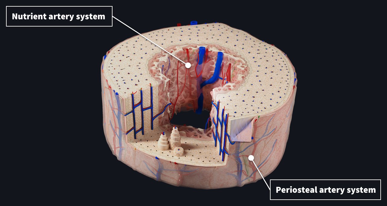
In addition to the mechanical functions of maintaining the body’s shape, allowing movement, and protecting internal organs, the skeleton is also the place of blood circulation: new blood cells are formed in the bone marrow (Luxgrant, 2020). Figure 3 shows the blood supply to a bone. The skeletal system is also involved in mineral metabolism: bones are a collection point for mineral salts (mainly calcium and phosphorus) necessary for bone formation (Luxgrant, 2020). It is also necessary for the functioning of the nervous system, muscles, blood clotting system, and other body systems.
Diseases
Many skeletal system diseases are known today; most are accompanied by mobility restriction, and some can lead to complete immobilization of a person. Malignant and benign bone tumors pose a severe threat to life and health and often require radical surgical treatment; the affected limb is usually amputated (I’Escalopier, Mathieu, Anract, & Biau, 2021). In addition to bones, joints, muscles, and connective tissues are often affected. Joint diseases are often accompanied by significant impairment of mobility and severe pain. Additionally, skeletal system diseases can often lead to complications in other bodily systems.
Summary
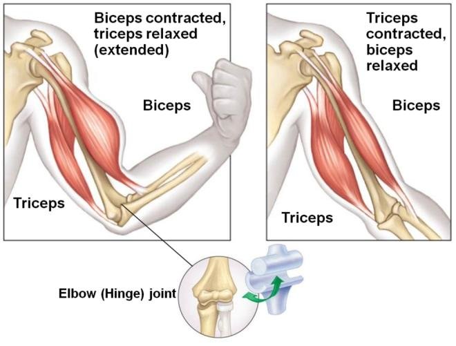
The skeletal system, comprising bone and cartilaginous elements, as well as associated connective tissue structures (such as ligaments), forms the passive component of the musculoskeletal system. That is complemented and driven by the skeletal muscles, the active part. Figure 4 is an example of how the human musculoskeletal system works. The spine, which holds the trunk upright and supports the skull, is essential (Figure 2). It is also the link between the chest and pelvis bones. The skeletal system supports the body; as a result, it supports the body while protecting critical internal organs. The human skeleton also serves as an attachment point for muscles that enable movements such as walking or raising an arm.
Before birth, the bone still consists of soft tissue; later, cartilage is formed from it, and gradually the bones become hard. The skeleton’s bones are involved in the processes of hematopoiesis and mineral metabolism, and the bone marrow is an essential part of the body’s immune system (Figure 3). In addition, the bones that make up the skeleton help the organs and soft tissues of the body and protect vital internal organs.
The skeletal system’s diseases affect the bones and joints and may damage the muscles or connective tissues. It can be an inflammatory, pathological, tumor, or other condition. Most often, they occur as independent diseases, but sometimes they can be symptoms of other diseases. Thus, the skeletal system is one of the most essential parts of a person’s existence and functioning. It has connections with other body systems, and diseases of this system can become life-threatening.
References
Dunn, P. (2022). Skeletal system functions & structures.
I’Escalopier, N., Mathieu, L., Anract, P., & Biau, D. (2021). Management of musculoskeletal tumours of the extremity in low-resource settings. International Orthopaedics, 46(2), 371-379.
Jones, K. E., Gonzalez, S., Angielczyk, K. D., & Pierce, S. E. (2020). Regionalization of the axial skeleton predates functional adaptation in the forerunners of mammals. Nature Ecology & Evolution, 4(3), 470-478.
Kan, H., & Strouse, P. J. (2015). Embryology, anatomy, and normal findings.
Luxgrant, A. (2020). Blood supply to the bone: Complete anatomy.
Mouath, A.M., Alzahab, N.A., & Alimam, H.S. (2017). Design of EMG Acquisition Circuit to control an antagonistic mechanism actuated by pneumatic artificial muscles PAMs.