Introduction
In this paper, the structures, systems, and organs of the skeletal and muscular systems and the digestive system will be discussed. This paper will review the main components of these systems and discuss how they aid the functioning of the human body. Additionally, for each system, two common diseases will be discussed and analyzed. The basis of this paper is the textbook by Tucker and internet sources that provide reliable information about human anatomy. The goal of this paper is to present an overview of the digestive and musculoskeletal systems and discuss the various elements of them.
The Skeletal and Muscular System; Structures, Systems, and Organs
The first section will discuss the structure and the functions of the skeletal system. There are 206 bones in the human skeleton, which are connected to one another through joints. There are three types of the latter: fixed, hinged, and ball and socket (Tucker, 2015). Diagram 1 is an illustration of the main bones that humans have with labels. The following sections will discuss in detail the elements of the skeletal system and the composition of the bones.
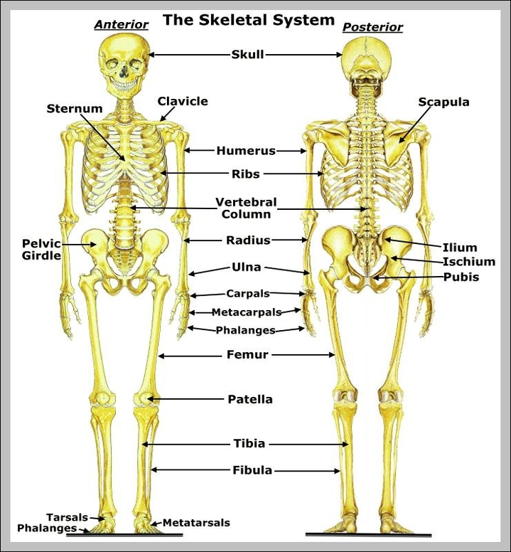
Among the functions of the skeletal system, there are the protective, supportive, movement, attachment, blood cells generation, as well as the storage of minerals and chemical energy storage (Tucker, 2015). Hence, apart from the skeleton supporting the human body and enabling its movement, which is achieved through the provision of the solid structure and the attachment of the points for the majority of the skeletal muscles, there are also other important contributions of this system to the human body’s ability to function. Additionally, the rigid structure of the bones allows them to protect the organs; for instance, vertebrae serve as a shied for the spinal cord (Tucker, 2015). The large bones contain the red bone marrow, where the blood cells are produced (Tucker, 2015). Finally, the essential minerals, such as calcium and phosphorus, are stored in the bone’s tissue, while the yellow bone marrow has fatty cells that are the body’s energy reserve (Tucker, 2015). Therefore, the skeletal system enables the diverse set of functions essential for the human body.
The main bones of the appendicular skeleton are the shoulders, hips, arms, and legs (Tucker, 2015). This structure consists of 80 bones, and all of the upper body bones belong to this category. The bones of the axillar skeleton are the vertebral column, ossicles, hyoid, sternum, and ribcage (Tucker, 2015). These bones are located in the head and vertebrate. The axial and the appendicular skeleton structures are the two categories used to classify the different types of bones in the human body.
The next paragraph will discuss the composition of the bones, which changes during the development stages of a person from an embryo to a grown person. According to Tucker (2015), embryos do not have rigid bones, and instead, the composition of this system is cartilage. This structure is firm, however, not as firm as the actual bones, and has elasticity (Tucker (2015). The composition of cartilage is collage and stretchy fibers, which allow for the elasticity of these structures. The deposition of calcium phosphate in the bones is what enables the transformation from cartilage to hard bones (Tucker, 2015). A developed human body’s bones are composed of calcium phosphate and fibers, and the latter allows the bones to remain unshattered upon not strong impacts. If the bones were to consist only of this mineral, they would shatter very easily, which would hinder the protective function of this system.
Apart from the collagen, fibers, and minerals, the bones’ tissue structure should be examined. There are three main layers: periosteum-sound, compact, and spongy (Tucker, 2015). The first element is a membrane that is necessary to protect the bone if there is no cartilage. Moreover, it contains the osteoblast-sound, which are also known as bone-forming cells (Tucker, 2015). The compact bone is what allows these structures to be hollow, and it houses the nerves. The final element is the hollow part of the bone that is located towards the end of these structures. There, the red marrow that produces blood cells is contained.
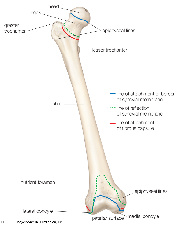
A long bone is a classification describing a structure that is longer in comparison to its width. Such bones have a shaft and may have several endings (Tucker, 2015). One example of a long bone is a femur, which is located in the upper part of the leg. The structure of a long bone in a human body is presented as Diagram 2, which shows the femur’s main elements. In this diagram, one can see the femur’s head, neck, shaft, and endings, which are the typical elements of a long bone.
The joints, as was mentioned, allow for the different bones to interconnect. The main joints in one’s body are: “fibrous, cartilage, and synovial” (Tucker, 2015, p. 50). Fibrous joints do not allow for movement, and their function is merely connecting the different parts of the body’s skeleton. The more flexible joints are the ones that are attached through cartilage. The synovial joints are the ones that allow the movement of the bones, and their structure contains fluid that allows to lubricate and safeguard the bones.
The main muscles of the body are classified into three categories, skeletal, cardiac, and smooth (Tucker, 2015). The contraction of the skeletal muscles allows the body to move, and these muscles are also categorized as voluntary because their movements are under a person’s conscious control. The cardiac muscles are essential for the cardiovascular system because they allow for the heart’s contraction and, therefore, enable the pumping of blood. These are the involuntary muscles because a person does not have conscious control over them (Tucker, 2015). The smooth muscles are also involuntary and can be found in the organs of the body. The functions of the muscles are evident, as their contraction allows for the body’s movement (Tucker, 2015). However, the cardiac muscles also enable blood circulation, while involuntary muscles in the inner walls of the organs help the latter contract. As for the structure, all muscles consist of blood vessels, fibrous tissue, and nerves (Tucker, 2015).
Two Diseases of the Skeletal System
The first disease that may affect the human body’s skeletal system is rickets. This condition is caused by dietary deficiency when a person does not consume sufficient amounts of calcium. Due to this, the bones become less rigid and more flexible, which means that they can no longer support the body (Tucker, 2015). Vitamin D deficiency also causes the development of rickets, and it is also linked to dietary deficiencies. Apart from this, Tucker (2015) notes that women are more susceptible to the development of this illness after menopause when compared to men. The second disease of the skeletal system is osteoporosis, which is a group of illnesses. According to Pessler (2020, para. 1), it is “a group of rare hereditary disorders that increase the density of bones and cause bones to grow abnormally.” As a result, an affected person has thicker bones which can easily break.
The two diseases that impact the muscular system are multiple sclerosis and polymyositis. Polymyositis is a rare category of myositis, which causes the inflammation of the muscle tissue and the blood vessels (Barhum, 2021). The main symptoms of this condition are weakness and inflammation of the muscles, dry cough, and issues with swallowing. Multiple sclerosis is a rare condition that is linked to the dysfunction of the neurons, and it affects the neurons in the spinal cord and the brain (Barhum, 2021). People with this condition lose the ability to control the voluntary muscles, and there is no treatment to prevent or reverse the effects of it, although some outcomes can be controlled and slowed with the use of medication.
The Digestive System
The digestive system enables the human body to use the nutrients that people consume with food and employ these elements either as energy or as the building blocks for the tissues. The organs in this system are classified into two categories: the main and accessory, with the former, also referred to as the alimentary canal (Tucker, 2015). The main organs are the ones that allow digesting the food, while accessory ones are needed to support this function.
Structures, Systems, and Organs
The main digestive system consists of the organs that facilitate the processing of the food from the moment it enters the human body until the remains that do not have nutritional value for the body leave it. There are six organs in the main digestive system: “mucosa, submucosa, muscularis externa, and serosa” (Tucker, 2015, p. 75). Notably, according to Tucker (2015), although these layers are present in all parts of the digestive system, the way they appear may differ significantly. Each element consists of its own structures; for example, the mucosa has epithelium, lamina propria, and muscularis mucosa.
The accessory organs of the digestive system are needed to support the functions of the main ones. For example, the teeth aid at the initial stages of food processing due to them helping break down the large chunks of the food into smaller ones. Other accessory organs include the granular organs and the tongue, all of which are required to shift the food from the mouth into the other parts of the digestive system.
The organs of the alimentary canal include the mouth, pharynx, esophagus, pharynx, small intestine, and large intestine (Tucker, 2015). The digestion process begins in the mouth or the oral cavity, where the food enters the human body. The process consists of chewing the food, which is the mechanical digestion, accompanied by saliva that allows for the food to be digested on a molecular level. Next, processed food enters the esophagus through the pharynx, the main function of which is to serve as a pathway from the oral cavity to the other parts of the digestive system. In the esophagus, the food passes through with the force of the muscle action (Tucker, 2015). The stomach, which is the next element of the digestive process, has an acidic environment. Due to this, the large molecules of food are broken down into smaller ones. This action is required to ensure that these molecules are processed and absorbed in the smaller intestine. In the small intestine, villi help absorb these molecules and the nutrients into the bloodstream. The cecum is the final part of the digestive system, where the remains of the food are processed as waste. There, the remnants of water from this waste are absorbed, and the remaining disease is moved to the rectum, which is a temporary storage space for them and into the anus. This final stage of the digestive process is also important because it allows the body to get rid of the food parts that could not be processed or which do not contain the nutrients it needs. The increasing pressure in the rectum forces the fecal matter through the rectal canal (Tucker, 2015). Thus, the process of digestion is complex, and over the course of it, the food is mechanically and chemically processed, and the organs of the digestive tract absorb the nutrients while the waste leaves the body.
Evidently, the goal of the digestive process is to allow the body to absorb the nutrients from the food consumed by an individual. Tucker (2015, p. 78) defines absorption as the “absorption is the movement of molecules across the gastrointestinal (GI) tract into the circulatory system.” The primary nutrients absorbed by the body are carbohydrates, fats, and proteins, as well as minerals and vitamins (Tucker, 2015). After the food is moved to the small intestine, the walls of this organ absorb the nutrients into the blood, where those are distributed across the body. Each of the main nutrients is broken down into smaller parts; for example, carbohydrates become simple sugars, while fats are transformed into fatty acids and glycerol, and protein becomes amino acids. Water is absorbed from the food as it is, and it is later used to provide fluids for the body.
As for the chemistry of digestion, each nutrient is broken down into a form that can be either immediately used by the human body or is transported into storage, from where it can be later used. For example, polysaccharides are broken into monosaccharides, for example, glucose. Next, through facilitated diffusion, these simple sugars are moved across the bloodstream. The proteins are broken down by peptides and pancreatic trypsin (Tucker, 2015). Next, the “fragments are then digested to free amino acids by carboxypeptidase from the pancreas and aminopeptidase from the intestinal epithelium” (Tucker, 2015, p. 80). After this, similar to monosaccharides, the amino acids are transported through facilitated diffusion across the bloodstream. The fats are processed with the help of pancreatic lipase. Emulsification allows the transformation of the large lipids into smaller ones. Hence, the chemistry of digestion relates to the acidic environment of the intestine, which allows the processed food and absorbs the molecules of nutrients.
Two Diseases of the Digestive System
Among the common diseases of the digestive system, the notable ones are acid reflux and gastritis. The former is a condition when what is contained in the stomach does not move towards the other organs of digestion, and instead, they are returned to the esophagus (Tucker, 2015). The symptoms of this condition can be repeated over time and lead to some complications, for example, causing heartburn. One cause of this illness is the relaxation of the sphincter muscles or other diseases that cause it to become weak. However, only lifestyle changes, such as diet, can help treat this condition. Next, gastritis is a more severe illness that affects the digestive system and can take a chronic form. When a person is affected by this illness, their mucosa is inflamed, causing damage to the stomach lining. This can lead to the inability to digest food or even bleeding.
Conclusion
In summary, this paper reviews the main structures of the muscular and skeletal, and well as digestive systems. Both are essential to the human body because the former allows for the movement and protection of the inner organs, while the latter enables the body to process foods into nutrients, which are later used as energy or building blocks for the cells or stored for the future. Both systems can be affected by a variety of diseases; for example, multiple sclerosis impacts the ability of the person to control their voluntary movements. Osteoporosis causes the unusual growth of the bones and causes them to be susceptible to breakage. Gastritis causes the inflammation of the stomach’s inner lining, while acid reflux occurs when the unprocessed food returns into the esophagus. This paper was completed using the textbook by Tucker and internet sources, and the presented diagrams outline the main elements of the musculoskeletal system and the structure of a large bone.
Reference list
Barhum, L. (2020) What are muscular system diseases? Web.
Femur (no date) Web.
Pessler, F. (2021) Osteopetroses. Web.
Tucker, L. (2015) An introductory guide to anatomy & physiology. London: EMS.
Skeletal system images (no date) Web.