Introduction
Crucial in achieving optimal benefits for all screening programs, quality assurance is defined as a system of procedures, checks, audits, and corrective measures to ensure that health services are at their highest achievable quality. Mammography is a screening and diagnostic tool which uses low-dose amplitude x-rays to examine the human breast, usually for the detection of breast cancer. The goal of a mammography exam is to detect very small abnormalities in the breast tissue before they develop into breast cancer (Saunderson, 2009). While quality assurance refers to systematic action in general, quality control refers to technical aspects alone. To ensure effective quality assurance and control, teamwork is needed. Overviews of existing quality assurance protocols in mammography will be outlined and contrasted with each other, with emphasis put on possible areas of improvement in the future. A questionnaire was sent to King Khaled General Hospital in Saudi Arabia to gather information regarding equipment management and use, including the presence of and use of a quality assurance program and the degree and techniques used to minimize radiation risk and provide user/patient safety. Using information provided through this questionnaire and also though later personal communications by Sultan Aawdh, medical physicist of the Radiological Department of King Khaled General Hospital in Saudi Arabia, this report describes and discusses the Quality Assurance/Control program for mammography used in King Khaled General Hospital in Saudi Arabia.
Description of Equipment Used for Mammography at the Radiological Department at King Khaled General Hospital in Saudi Arabia, the Quality Assurance Program within this Department and Existing National Standards
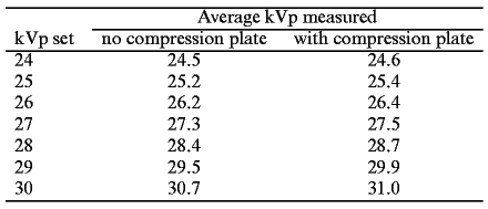
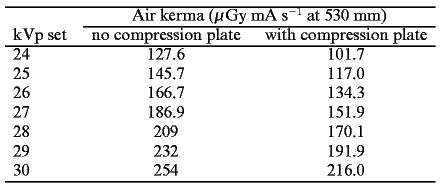
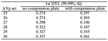
According to the initial information gained from the questionnaire provided to Aawdh, the Radiological Department at King Khaled General Hospital in Saudi Arabia uses the Mammomat-2 machine, manufactured by Siemens, for mammography exams. The current machine in use was installed in 1991. This machine was supplied together with a machine guidance manual. The Measured characteristics of the Siemens Mammomat 2 is Table 1 give the measured kVp average, both with and without the compression plate in situ. Table 2 gives the measured air kerma, both with and without the compression plate in situ. The compression plate absorbed 20.3% of the primary beam at 24 kVp (set) and 15% at 30 kVp (set). Table 3 gives the measured half value layer, both with and without the compression plate in situ. The coefficient of variation for both kilovoltage and air kerma was less than 0.05 (over 10 measurements) (Hartley, 1999).
Aawdh stated on the questionnaire that all requirements are in place to improve the quality of the machine, prove the image quality and quality control measures. He states, a weekly compression and visual test are conducted weekly, and QC tests for output and KVp are carried out annually. Also he stated that as part of the maintenance and quality assurance schedule for the machine a schedule for P.P.M. is done every 8 months. Furthermore, he stated that the following is done to improve the quality of images. Firstly, the department ensures that their machines are in good condition and that all necessary tests are carried out. Secondly, appropriate exposure factors and good position are maintained. Finally, the department aims not to repeat any procedures. In response to information regarding the radiation safety program followed by the department, Aawdh mentions 2 principles that are followed for radiation safety program. These are: Justification for use. Mammography is carried out routinely on females starting from age 45 and patients are only examined once a year unless recommended otherwise by a doctor. The next principle that is followed is optimization. This relies on the principle of ALARA, which he describes as getting the best image using the least exposure, and not repeating procedures unnecessarily. It was discovered that the Radiological Department at King Khaled General Hospital in Saudi Arabia follows the standards as required by the Saudi Ministry of Health under Section 900.12(d) of the Mammography Quality Standards (Aawdh, personal communication 26, April 2010). This Standard makes the following specifications. Firstly, the facility shall designate a lead interpreting physician. Secondly, the lead interpreting physician and department director will assign a quality control technologist. Thirdly, a medical physicist shall be assigned to work with the lead interpreting physician and quality control technologists to establish and maintain the program. Finally, each individual will be responsible for specific components of the Mammography Quality Assurance Program as defined in the Mammography Quality Assurance Responsibility Policy (Aawdh, personal communication 26, April 2010). In addition, this Standard sets out who is responsible for the Quality Assurance Program. That is a Lead Interpreting Physician is designated the lead interpreting physician and is responsible for overseeing mammography quality and ensuring compliance with the Mammography Quality Standards and the Rules of Good Practice. While a Quality Control Technologist is designated as quality control technologists and is responsible for all quality control tests and records maintenance not performed by the physicist or interpreting physician. Further, a Medical Physicist will act as medical physicist and is responsible for all annual QC testing and for overseeing all mammography equipment. Furthermore, Section 900.12 (e) of the Mammography Quality Standards, in accordance with the Mammography Quality Control Manual of the American College of Radiology, sets out the requisite equipment quality control tests. This section sets out the tests are required when. For example, Processor Quality Control tests must be carried out daily, Image Quality Evaluation tests must be done weekly, Fixer Retention and Repeat Analysis done every 3 months, Darkroom Fog, Screen-Film Contact, and Compression done 6 monthly, while Automatic Exposure Control, KVp Accuracy, Focal Spot size or resolution, Half Value Layer, AEC Reproducibility/Breast entrance air kerma, Average Glandular Tissue Dose, X-ray Field/Light Field/Image Receptor Compression paddle Alignment, Uniformity of Screen Speed, System Artifacts Radiation Output Decompression must be carried out every 12 months. The Standard also specifies that mammography exams cannot be performed if the following Quality Control tests fall outside of control limits: Processor QC, Phantom Image Test, Darkroom fog, Screen film contact, Compression Test, or Average glandular dose. As for all other Quality Control tests that fall outside of control limits, these must be corrected within 30 days as defined in Section 900.12 (e)(8)(ii)(A) of the Mammography Quality Standards (Aawdh, personal communication 26, April 2010).
Discussion
Radiation Safety Implications of Mammography Equipment for Patients
The dose of radiation from mammography might seem high when compared to other X-ray exams; however, the patient’s overall risk from a single mammography exam is quite low (Saunderson, 2009). It is important for medical practitioners to remember that mammographic film produced by a radiation dose that is too low can also be harmful (Saunderson, 2009). An image of the breast formed by a very low dose of radiation does not have high image quality as compared to an image produced using an adequate radiation dose which is established by a medical physicist evaluation and a good quality assurance program. One of the most important image qualities in mammography is the level of contrast. High contrast is necessary to detect microcalcifications that may occur in a pre-stage of cancer. As the level of KVp (kilo volt peak) decreases, the contrast increases due to the attenuation coefficient differences between tumor tissue and normal soft tissue of the breast. In mammography, medical practitioners are sometimes tempted to use a lower KVp in order to increase contrast. With a lower KVp, more radiation is retained within the body, so the dose goes up. In order to decrease the dose of radiation that enters a patient’s body during mammography, one has to increase the amount of KVp that is used (Harris, 2000).
Radiation Safety Implications of Mammography Equipment for Staff
A room where mammographic X-ray examinations are done must not be used for more than one radiological investigation simultaneously. Except for those persons whose presence is essential for the investigation, all other persons must leave the room when a mammographic X-ray examination is carried out. Personnel must, at all times, keep as far away from the radiation beam as practicable. Radiation exposure of personnel by the primary X-ray beam must never be allowed unless the beam is adequately attenuated by protective screens or protective clothing. Deliberate irradiation of an individual for training purposes or equipment evaluation must never occur and all personnel must take full advantage of available protective devices. Operation of the X-ray tube should be controlled from the control panel which is usually located behind a protective screen or inside a control booth. The technologist must be shielded when exposures are made. If a patient escort or other person is called upon to assist in any situation, the person must be provided with protective clothing and be positioned so as to avoid the primary X-ray beam. No one must regularly perform these duties. All technologists operating X-ray equipment and who are likely to receive a radiation dose in excess of 5 percent of the recommended dose limits for radiation workers must wear personnel dosimeters. All entrance doors to a mammographic X-ray room, including patient dressing room doors, must be kept closed while a patient is in the X-ray room. There should be a light or a sign on the outside of the patient dressing room indicating occupancy (Health Canada, 2008).
Evaluation
Quality Control is an evaluation program of both the equipment and processes used to carry out a given activity or operation. In general, the QC program at the hospital appears adequate for the following reasons. Firstly, (Bushong, 1997, 306) states that QC involves testing, record keeping together with the evaluation of the imaging equipment and the processing of the image. He also maintains that within a workplace, the radiographer, medical physicist, and QC mammographer must all work as one in this evaluation (Bushong, 1997, 306). As described above, it can be seen that in the case of King Khaled General Hospital, such a program is indeed in place. For instance, the national Standard adopted by the hospital specifies the required tests together with the timing of these tests, and also the responsibilities for each team member have clearly been specified.
Secondly, there appears to be a high standard of QC measures since the hospital has adopted standards that are based on national legislation as set by the Ministry of Health. In addition to these, these national standards are based on guidelines set by the Mammography Quality Control Manual of the American College of Radiology, and the American College of Radiology (ACR) is recognized around the world in the field of radiology.
While there are clear and reliable standards in place for the hospital which indicate that a good QC program is in place, there are some other factors that could be considered. For example, the hospital could consider adopting a random on-site survey as is required by the FDA in America of all mammography centers in the USA. However, it must be kept in mind that while in an ideal world the mammography would be made to conform to the safest of all possible standards, it is usually not a feasible project that can be implemented in the real world. This is because, costs and time allowed for QC and QA must also be considered when designing and implementing programs for mammography.
Conclusion
In conclusion, this report has used information gained from a questionnaire and personal communication with a medical physicist at the King Khaled General Hospital in Saudi Arabia to describe the Quality Assurance (QA) and Quality Control (QC) programs in place at the hospital. These QA and QC programs indicated that the management of the hospital wanted to protect the health of patients while ensuring that the staff of the hospital is safe against infections during mammography. The programs were evaluated as being effective and sufficient since they conformed to the standards and guidelines on QC and QA programs as set out by the Ministry of Health in Saudi Arabia and they were based on standards established by the Mammography Quality Control Manual of the American College of Radiology. However, it is generally acknowledged that King Khaled General Hospital has well established QA and QC programs but they are not regularly checked to assess their performance. Due to this, one factor that could be adopted from abroad is the use of spot checks from an external body to ensure that patients and staff at this hospital enjoy the best possible outcomes. This will make the management of the hospital to ensure that the QA and QC programs always conform to the desired standards.
References List
Bushong, S. C. 1997. Radiologic Science For Technologists Physics, Biology, and Protection 6ed. Texas: Mosby.
Harris, C. H. 2000. Quest for Quality: QA and QC in Medical Imaging. Imaging Economics. Web.
Health Canada. 2008. Radiation Protection in Mammography: Recommended Safety Procedures for th Use of Mammographic X-Ray Equipment.
Hartley, L. D., B. J. Cobb, and D. E. Hutchinson. 1999. Estimating mean glandular dose using proprietary mammography phantoms. Applied Radiation and Isotopes 50 (1): 205-213. Web.
Saunderson, J. 2009. Radiation Protection for Assistant Practitioners Mammography:Lecture4. Web.