Endomitosis in Drosophila Virilis

Background & Purpose

Endomitosis is a variant of the cell cycle that leads to the production of up to a thousand copies of the genome. During this process, the cell does not divide but the chromosomes increase in size as the number of copies of genes increases. The purpose of this lab was to investigate endomitosis in the salivary glands of Drosophila virilis larvae. It was hypothesized that endomitosis also occurs in the salivary glands of Drosophila virilis. This hypothesis was formulated based on the fact that endomitosis has been reported to occur in Drosophila melanogaster, which is closely related to Drosophila virilis.

Results

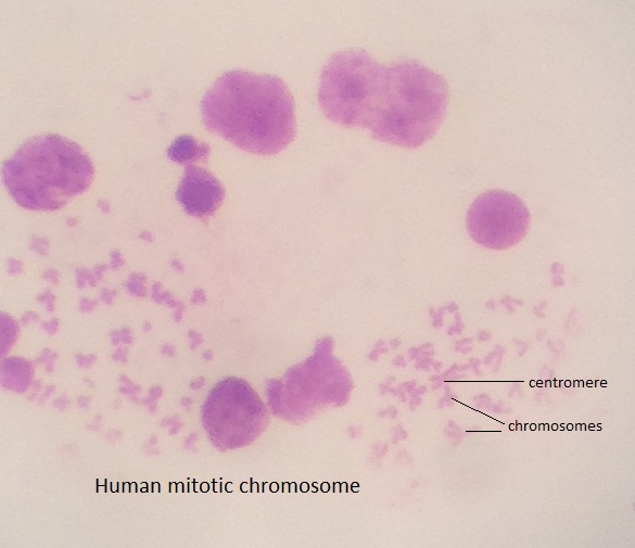
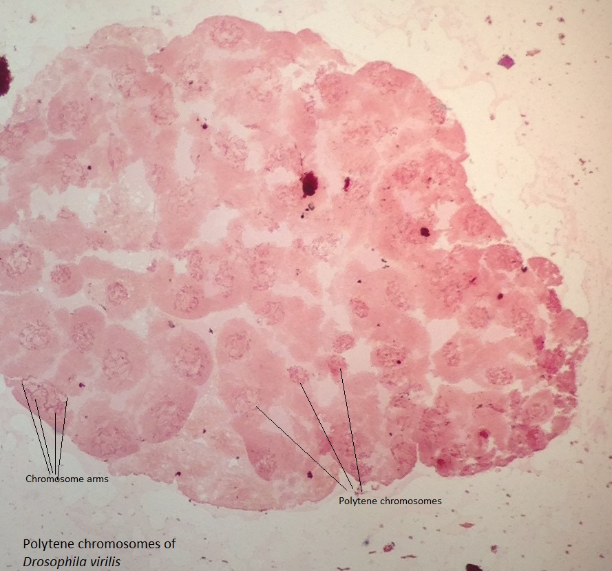
The images of the human cells and salivary glands of Drosophila virilis as observed under the compound microscope are shown in the appendix in figures 1 and 2.

Discussion

It was hypothesized that endomitosis took place in the salivary glands of Drosophila virilis. The findings of the experiment showed the presence of polytene chromosomes, which were only produced in endomitosis, in the salivary glands of the larvae of Drosophila virilis as shown in figure 2. The polytene chromosomes were distinctive from the mitotic chromosomes in human cells (shown in figure 1). Some of the major differences were the sizes of the two chromosomes. Figure 1 showed small mitotic chromosomes in human cells while figure 2 showed significantly big polytene chromosomes in the salivary glands of Drosophila virilis. Another major difference was the presence of several polytene chromosome arms in Drosophila virilis whereas the human mitotic chromosome only had four distinctive arms. However, the chromocenter of the polytene chromosome was not easily distinguishable. Therefore, the findings of this experiment supported the hypothesis that endomitosis occurred in the salivary glands of Drosophila virilis larvae.

Appendix

Figures

Mitotic chromosomes in human cells
Figure 1: Mitotic chromosomes in human cells. The above figure shows the mitotic chromosomes in humans as well as distinct chromosomes. The samples were obtained by staining the cells in aceto-orcein stain and observing under a compound microscope at ×40 objective lens.
Polytene chromosomes of Drosophila virilis.
Figure 2: Polytene chromosomes of Drosophila virilis. The above figure shows the polytene chromosomes in Drosophila virilis as well as the chromosome arms. The samples were obtained by staining the salivary glands of D. virilis in aceto-orcein stain and observing under a compound microscope at ×40 objective lens.

Cite this paper

Select style

Reference

StudyCorgi. (2022, June 13). Endomitosis in Drosophila Virilis. https://studycorgi.com/endomitosis-in-drosophila-virilis/

Work Cited

"Endomitosis in Drosophila Virilis." StudyCorgi, 13 June 2022, studycorgi.com/endomitosis-in-drosophila-virilis/.

* Hyperlink the URL after pasting it to your document

References

StudyCorgi. (2022) 'Endomitosis in Drosophila Virilis'. 13 June.

1. StudyCorgi. "Endomitosis in Drosophila Virilis." June 13, 2022. https://studycorgi.com/endomitosis-in-drosophila-virilis/.


Bibliography


StudyCorgi. "Endomitosis in Drosophila Virilis." June 13, 2022. https://studycorgi.com/endomitosis-in-drosophila-virilis/.

References

StudyCorgi. 2022. "Endomitosis in Drosophila Virilis." June 13, 2022. https://studycorgi.com/endomitosis-in-drosophila-virilis/.

This paper, “Endomitosis in Drosophila Virilis”, was written and voluntary submitted to our free essay database by a straight-A student. Please ensure you properly reference the paper if you're using it to write your assignment.

Before publication, the StudyCorgi editorial team proofread and checked the paper to make sure it meets the highest standards in terms of grammar, punctuation, style, fact accuracy, copyright issues, and inclusive language. Last updated: .

If you are the author of this paper and no longer wish to have it published on StudyCorgi, request the removal. Please use the “Donate your paper” form to submit an essay.