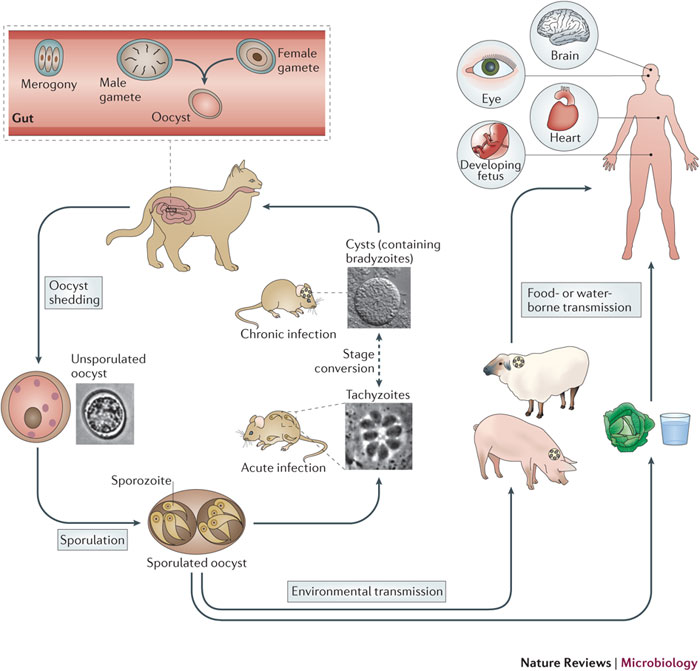
Comparison between tachyzoites and bradyzoites
During the various stages of a lifecycle, a parasite goes through various cellular stages that are characterized by different morphology, behavior, function, and biochemistry. Tachyzoites and bradyzoites are stages in the lifecycle of T. gondii. In each of the stages, the parasite differs in shape, size, function, and location. The main function of tachyzoites is to expand the population of the parasite in the host through rapid multiplication (Hill & Dubey 2002). Their motility and ability to multiply rapidly aid in fulfilling their role. With regard to their morphology, tachyzoites are crescent-shaped and possess a pointed front (Dubey & Jones 2008). In addition, they are 2 by 6 micrometers with a rounded back end. They contain numerous organelles and inclusion bodies. Examples of these structural bodies and organelles include pellicle, micronemes, endoplasmic reticulum, apical rings, Golgi complex, ribosomes, microtubules, amylopectin granules, micropores, mitochondria, and dense granules (Dubey 1998). They lack motility structures even though they have the ability to flex, glide, undulate, and rotate. After multiplication, they are transported to various parts of the body through bloodstreams. As the lifecycle progresses, tachyzoites convert to bradyzoites in order to form tissue cysts that are critical in the development of the parasite (Eaton 2014). Unlike tachyzoites which have a central nucleus, bradyzoites have a nucleus that is located toward the cell’s posterior end (Dubey 1998). They are crescent-shaped and are larger than tachyzoites. They are about 7 by 1.5 micrometers in size. The rhoptries of bradyzoites contain numerous electrons while those of tachyzoites are labyrinthine (Offenberg 2015). The main function of bradyzoites is to form tissue cysts when they enter a host’s cells and aid in the progression of the parasite’s life cycle. They are orally infectious and therefore, play an important role in the transmission of T. gondii in the host’s body (Tenter et al. 2001).
Host’s immune response to Toxoplasma gondii
The host responds to infection by initiating innate and adaptive immune responses. After the ingestion of T. gondii, the host’s immune system activates macrophages to fight the parasite (Flegr 2013). The main purpose of the innate immune response is to prevent the multiplication of T. gondii (Tenter et al. 2001). In addition, it initiates the activation of the adaptive immune response after the ingestion of the parasite (Flegr 2007). The adaptive immune response triggers the release of certain antibodies and effector cells whose role is to eliminate the invader (Flegr 2013). The response causes the specialization of dendritic cells, B cells, and macrophages in order to present specific antigens for the elimination of T. gondii. The presentation of the antigen to T cells commences the differentiation process that leads to the development of immunological memory that protects the host from re-infection (Blanchard et al. 2015).
Localization of the cysts on the brain
One of the mechanisms through which T. gondii manipulates the host’s behavior is through localization on certain parts of the brain. In infected hosts, cysts of the parasite are usually distributed in several brain regions (Carruthers & Suzuki 2007). A study conducted to study the distribution of T. gondii cysts in the brains of CD1 mice found out that cysts were localized on all brain regions including the olfactory bulb, the hippocampus, amygdala, the entorhinal, and the frontal association and visual cortices (Berenreiterova et al. 2011). Low distribution of cysts was observed in regions that include the cerebellum, myelinated axons, the pontine nuclei, and the caudate-putamen. The study conducted by Berenreiterova et al (2011) found out that 54 brain regions contained the parasite’s cysts. Research has shown that during the chronic stages of T. gondii infection, cysts are found throughout the brain (Blanchard et al. 2015). However, the localization of the cysts has not yet been studied in detail. Certain studies have shown that there is a high density of T. gondii cysts in two main brain regions namely the frontal cortex and the amygdala (Carruthers & Suzuki 2007). Brains infected by T. gondii have high volumes of dopamine (Lafferty 2006). These findings have been used to explain why infected hosts exhibit changes in behavior. According to McConkey et al (2013), T. gondii manipulates host behavior by localizing in brain regions that process fear including the amygdala (Wiser 2010). It can be concluded from several research studies that T. gondii cysts are highly localized in brain regions that include the amygdala, frontal cortex, olfactory bulbs, hippocampus, and diencephalon (McConkey et al. 2013).
Effect on neuromodulator levels
Studies have proposed histopathological, immunological, and neuromodulatory hypotheses for the manipulation of host behavior by T. gondii. According to the neuromodulatory hypothesis, the local immune response that is elicited to inactivate T. gondii alters the levels of cytokines, which influence neuromodulator levels (Lafferty 2006). The neurological basis of anxiety has been studied in several studies to determine how T. gondii alters neuromodulator levels. Fearless reactions in rats have been shown to emanate from the blocking of anxiogenic N-methyl-D-aspartic acid receptors in the amygdala (plays an important role in emotional behavior). Homovanillic and norepinephrine alter the mood, locomotor activity, and cerebral blood flow of hosts. Homovanillic acid is a degradation product of dopamine that has been associated with behavior change of infected hosts. One of the proposed mechanisms of neuromodulation involves alterations in the levels of neurotransmitters in the host (Wiser 2010). Brain cells of acutely-infected hosts show an HVA elevation of 140% while the levels of dopamine in chronically-infected hosts elevate by 114% (McConkey et al. 2013). Several studies have found that the levels of dopamine increase in brain cells that contain cysts. T. gondii directly increases the quantities of dopamine in infected cells by synthesizing tyrosine hydroxylase, which plays an important role in the production of dopamine. Studies have identified two tyrosine hydroxylase genes that are responsible for the production of excess dopamine in the brain. For example, overregulation of the TgAaaH2 gene during the differentiation of T. gondii to bradyzoites results in stimulation that increases the production of dopamine (McConkey et al. 2013). The accumulation of dopamine in various brain regions is responsible for the behavioral changes that are observed in animals and humans infected by T. gondii (Lafferty 2006).
References
Berenreiterova, M, Flegr, J, Kubena, A & Nemec, P 2011, ‘The Distribution of Toxoplasma gondii Cysts in the Brain of a Mouse with Latent Toxoplasmosis: Implications for the Behavioural Manipulation Hypothesis’, PLoS One, vol. 6, no. 12, 41-53.
Blanchard, N, Dunay IR & Schulter, D 2015, ‘Persistence of Toxoplasma gondii in the Central Nervous System: A Fine-Tuned Balance between the Parasite, the Brain and the Immune System’, Parasite Immunology, vol. 37, no. 3, 150-158.
Carruthers, V & Suzuki, Y 2007, ‘Effects of Toxoplasma gondii Infection on the Brain’, Schizophrenia Bulletin, vol. 33, no. 3, 745-751.
Dubey, JP & Jones, JL 2008, ‘Toxoplasma gondii Infection in Humans and Animals in the United States’, International Journal of Parasitology, vol. 38, no. 11, 1257-1278.
Dubey, JP 1998, ‘Advances in the Life Cycle of Toxoplasma gondii’, International Journal of Parasitology, vol. 28, 1019-1024.
Eaton J 2014, ‘What Does it Mean when 2 Billion people Share Their Brain with a Parasite’. Web.
Flegr, J 2007, ‘Effects of Toxoplasma on Human Behavior’, Schizophrenia Bulletin, vol. 33, no, 3, 757-760.
Flegr, J 2013, ‘Influence of Latent Toxoplasma Infection on Human Personality, Physiology and Morphology: Pros and Cons of the Toxoplasma-Human Model in Studying the Manipulation Hypothesis’, Journal of Experimental Biology, vol. 216, no. 1, 127-216.
Hill, D & Dubey, JP 2002, ‘Toxoplasma gondii: Transmission, Diagnosis and Prevention’, Clinical Microbiology and Infection, vol. 8, no. 10, 634-640.
Hunter, C & Sibley, D 2012, ‘Modulation of Innate Immunity by Toxoplasma gondii Virulence Effectors’, Nature Reviews Microbiology, vol. 10, no. 1, 766-778.
Lafferty, KD 2006, ‘Can the Common Brain Parasite, Toxoplasma gondii, Influence Human Culture?’, Proceedings of the Royal Society, vol. 273, no. 1602, 111-126.
McConkey, GA, Martin, HL, Bristow, GC & Webster, JP 2013, ‘Toxoplasma gondii Infection and Behavior: Location, Location, Location’, The Journal of Experimental Biology, vol. 216, 113-119.
Offenberg, K 2015, Toxoplasma gondii, Agenda Verlag GmbH & Company, New York.
Tenter, A. M, Heckeroth, A. R & Weiss, LM 2001, ‘Toxoplasma gondii: from Animals to humans’, International Journal of Parasitology, vol. 31, no. 2, 217-220.
Wiser, M 2010, Protozoa and Human Disease, Garland Science, New York.