Introduction
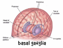
Cerebral palsy is a non-progressive condition that results from brain damage to the fetus during embryological development or immediately after birth (Carroll and Robert 468). Due to its early onset in a person’s life, the condition is diagnosed in infancy and early childhood. Clinical features of the condition are abnormal behaviors, posture abnormalities, epileptic seizures, nervous system disturbances, and problems in walking due to muscular tension. The use of stem cells to cure cerebral palsy involves the introduction of the cells to the affected brain region so that they could grow and enable the part of the brain to regain its normal physiological functions (Goldstein 45).
Literature review
In the developed world, every 2 in 1,000 births are affected by cerebral palsy whereby males are impacted to a greater extent than females across the world. There have been concerns that if the injuries to the brains of a fetus or infant are not minimized, cerebral palsy cases could be on the rise. Many countries have attempted to encourage expectant women to avoid exposing their fetuses in the womb to pressure that could lead to brain injury. In addition, women are encouraged to take care of their infants after birth so that they prevent them from brain injuries that could lead to cerebral palsy. There has been an agreement among scientists that cerebral palsy occurs when an injury to the developing brain interferes with the normal blood flow to the brain tissue (Goldstein 45). An injury to the cerebrum hinders normal blood and oxygen flow to the portion of the brain that is responsible for body movements. The injured cerebrum gets underdeveloped, and it does not perform its normal functions in the future (Goldstein 45).

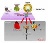
Stem cells have been shown to develop into any form of cells in the body based on the growth signals exposed to them (Carroll and Robert 468). Previous studies have identified how stem cells could be handled and used for clinical applications (Koning, Walton, Shin, Chen, et al 323). The goal of the use of stem cells in the cure of cerebral palsy is to replace the damaged tissue with cells that could develop into functional brain cells. The nervous system has been targeted for transplantation using stem cells because the system contains certain cell lines (Daadi, Davis, Arac, Li, et al 517).
Mesenchymal stem cells found in the bone marrow and umbilical cords migrate to the brain after an injury to initiate the repair of the damaged tissue. However, the cells provide only nutrients but cannot develop into mature neurons in animals. Neural precursor cells are found in the nervous system of a human being only. Some drug substances can activate the cells, which moves them towards the affected cerebrum where they initiate the repair of the damaged tissue. Pluripotent stem cells can develop into many cell types in the body. They could be utilized in the cure of cerebral palsy being converted to precursor stem cells which move to the brain to repair the damaged tissue (Daadi, Davis, Arac, Li, et al 517).
Methods
The methods described here were used in a previous study aimed to derive neural stem cells from a laboratory animal with a neural degenerative condition (Koning, et al 323). Young female and male transgenic mice were utilized to yield NSCs isolation. Older males were used for in vivo studies. Standard handling measures were used to avoid the contamination of the mice prior to the killing. Mutants and wildtype (WT) male and female mice were used. Their brains were isolated and processed appropriately to prevent disruptions of normal physiological functions. Genotyping was conducted using a 3-primer design PCR. Adherent cell cultures (NSCs) were grown on a neurobasal medium enriched with supplements. Growth factors were added to the media (to promote growth) and antibiotics (to prevent contamination). The NSC cultures were monitored regularly, and the cell cultures were stopped upon confirming that they had grown to the required numbers. To ensure continuous growth of the cells in the culture media, the growth media and other components essential for growth were changed every 3 days. The cultured cells were injected into the brain of mice with neurological disorders and monitored for some time. The cultured cells were also injected into the brain of the WT mice. Proliferative activities of progenitor cells were compared in WT and mutant mice. Immunohistochemistry (immunocytochemistry) assays were used to examine the neurological growth patterns of the mice which were injected with the cultured mutant and WT cells (Koning, Walton, Shin, Chen, et al 323). The overall goal of the assays was to determine the numbers of neurons initiated to grow and their rates of growth.

Results
The mutant neural basal cells produced more neurons at the beginning than the WT cultured cells. However, the mutant cells did not express adequate markers required for maturation. In addition, the neurons derived from the mutant cells showed abnormal morphologies and had higher levels of calretinin. The study also demonstrated that the neurogenesis induced by the mutant-derived cells was naturally programmed in mice, and was not a product of environmental manipulations (Koning, et al 323).
Discussion and conclusion
In vivo, both the WT and mutant mice produced many neurons that were initiated to grow by the cultured cells. However, the neurons did not mature to assume their physiological roles. The maturation markers were most impacted in vitro, but apoptosis was higher in the differentiating neurons induced by the mutant cells than in the neurons induced by the WT cells. It was concluded that the use of stem cells in the treatment of neurological disorders (including cerebral palsy) was an essential clinical step towards their management.
References
Carroll, James and Robert Mays. “Update on stem cell therapy for cerebral palsy.” Expert opinion on biological therapy 11.4 (2011): 463-471. Print.
Daadi, Marcel, Alexis Davis, Ahmet Arac, Zongjin Li, et al. “Human neural stem cell grafts modify microglial response and enhance axonal sprouting in neonatal hypoxic–ischemic brain injury.” Stroke 41.3 (2010): 516-523. Print.
Goldstein, Murray. “The treatment of cerebral palsy: what we know, what we don’t know.” The Journal of pediatrics 145.2 (2004): 42-46. Print.
De Koning, Eelco, Noah Walton, Richard Shin, Quingfeng Chen, et al. “Derivation of neural stem cells from an animal model of psychiatric disease.” Translational psychiatry 3.11 (2013): 323. Print.