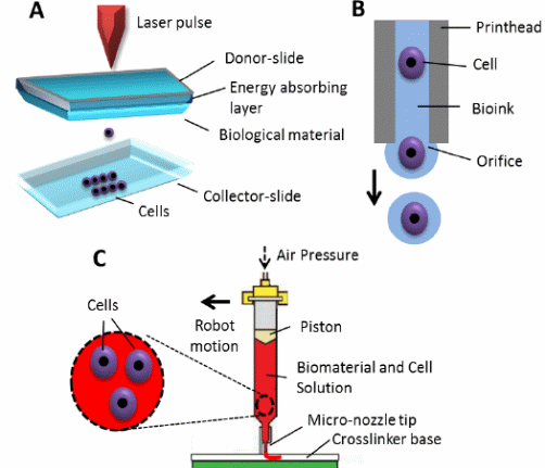
Bioprinting is a bioengineering process, in which Computer-Aided Design (CAD) and Computer-Aided Manufacturing (CAM) are used in biological fields such as creating artificial organs. Bioprinting is similar to 3D printing, as a 3D model is created by layer by layer deposition of certain material. Based on working principle, bioprinting can be classified into laser-based, inkjet-based and extrusion-based bioprinting (see fig. 1). Each one of these techniques has advantages and limitations. One of the most important applications of bioprinting is organ transplantation. Many promising trends are under research, offering hope for solving the problems that face these techniques.

Despite the great progress achieved in the field of bioprinting, there are still many challenges facing these techniques. Laser-based system has a limitation in printing in third dimension, inkjet-based system has a limitation of significant cell damage, and extrusion-based system has a limitation of shear stress induced cell deformation. Constructing hollow tubular and solid organs, constructing organs of high vascularity, sufficient mechanical strength and integrity of the constructs, fabricating 3D structures with high resolution, and developing biofunctional printable bioink materials with optimum properties are some of the obstacles that need to be overcome (Seol, Kang and Lee 347).
Researchers have been trying to overcome these difficulties, targeting to obtain fully functioning organs. Four-dimensional (4D) bioprinting is a recent technology that has the ability of shape transformation in response to internal or external stimuli. This technology paves the way for fabricating hollow self-folding tubes with capability to control their diameters and structural design at high resolution. Subsequently, this allows obtaining tubes of very small internal tube diameters, which are comparable to the smallest blood vessels (Kirillova, Maxson and Stoychev 7).
The bioprinting materials, bioinks, are natural materials, synthetic materials or hybrid biomaterials. Each one of these types has advantages and disadvantages. In order to mimic the natural microenvironment, a recent strategy is being developed. It depends on using cells that can produce natural extra-cellular matrix (ECM). The basis of this strategy is to seed the porous graft with cells that are able to secrete ECM, and then these cells are induced to apoptosis. The resulting devitalized graft can be stored until seeded with autologous cells (Arslan-Yildiz, El Assal and Chen 7).
Spheroid printing is a promising technique in biofabrication field. Spheroids are building blocks that do not need scaffolds. They are capable of self-assembly, shape maintenance, and in-place structuralization. Many researches are being conducted in order to use tissue spheroids to create a vascular tree. They are based on fabricating three types of spheroids: solid spheroids, mono-lumenized spheroids, and micro-vascularized tissue spheroids (Patra and Young 96).
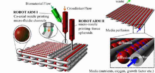
Spheroids have paved the way for the idea of organ printing. Organ printing is defined as a computer-aided process that necessitates the integration of three different types of technology: cell technology, biomanufacturing technology, and technologies for in vivo integration (Ozbolat and Yin 5). A conceptual model of 3D organ printing is related to tissue spheroids (see fig.2).

Fig.2. A conceptual model of 3D organ printing. Micro-fluidic channels (vessel-like) are printed in tandem with tissue spheroids, then connected to a bioreactor for media perfusion (Ozbolat and Yin 6).
Another promising trend in bioprinting is in-situ printing. It is based on intra-operational printing of living organs in the human body. This trend has already been tested for external organs such as repairing of a wounded skin, but it is still under research for internal organs. Miniature organs are considered a transition on the way to fully functioning organs. They are built in small scale, perform the most vital function, and are put in less immune-responsive sites. An example of printed miniature organ is the pancreas (Ozbolat and Yin 7).
In conclusion, bioprinting techniques have witnessed great advances in the last decades. They are expected to have more improvements to meet the increased need for organ transplantation. They offer a hope for managing many clinical problems that require transplantation as an appropriate line of treatment.
Works Cited
Arslan-Yildiz, Ahu, et al. “Towards Artificial Tissue Models: Past, Present, and Future of 3D Bioprinting.” Biofabrication, vol. 8, no.1, 2016, pp. 014103.
Kirillova, Alina, et al. “4D Biofabrication Using Shape-Morphing Hydrogels.” Advanced Materials, 2017.
Ozbolat, Ibrahim T. and Yu. Yin. “Bioprinting Toward Organ Fabrication: Challenges and Future Trends.” IEEE Transactions on Biomedical Engineering, vol. 60, no. 3, 2013, pp. 691-699.
Patra, Satyajit and Vanesa Young. “A review of 3D Printing Techniques and The Future in Biofabrication of Bioprinted Tissue.” Cell Biochemistry and Biophysics, vol. 74, no. 2, 2016, pp. 93-98.
Seol, Young-Joon, et al. “Bioprinting Technology and Its Applications.” European Journal of Cardio-Thoracic Surgery, vol.46, no.3, 2014, pp. 342-348.